Nervous system · Head and neck
Sensory cortex !
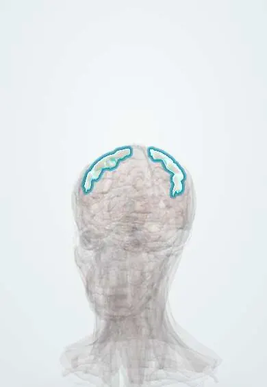
The sensory cortex (postcentral gyrus) is a strip of brain behind the central groove; it receives touch, pain, temperature and position from the opposite side.
Official name: Postcentral gyrus
Opens in the Durgical app, no sign-up needed.
What it is
The strip of cortex just behind the central sulcus.
What it does
Receives touch, pain, temperature and position from the opposite side of the body.
Key facts
- Where
- Directly behind the central sulcus
- Blood supply
- MCA (face, arm), ACA (leg)
- Function
- Primary somatosensory cortex (areas 3, 1, 2); opposite side
Relations
- In front
- Central sulcus, with the motor cortex (precentral gyrus) in front
- Behind
- Postcentral sulcus, then the superior parietal lobule and the supramarginal gyrus
- Below
- Lateral sulcus; the secondary somatosensory cortex lies on the parietal operculum, the upper bank of the fissure
- Medial
- Back of the paracentral lobule on the medial surface (leg, foot and perineum)
How to find it
On axial MRI it lies just behind the hand knob of the precentral gyrus and is thinner than it; the marginal ramus of the cingulate sulcus (bracket sign) reaches the top of the hemisphere just behind it. On the lateral surface the postcentral sulcus runs parallel to the central sulcus, behind it.
Examination
- Two-point discrimination on the fingertips: a larger gap is needed on the side opposite a cortical lesion
- Stereognosis and graphesthesia: failure to identify objects or traced numbers despite intact touch points to the parietal cortex
- Joint position sense: small up-or-down movements of a finger or toe are misjudged
On imaging
MRI shows it best. On axial images it is the gyrus just behind the central sulcus and its hand knob; the marginal ramus of the cingulate sulcus helps find it. An acute infarct is bright on diffusion-weighted imaging (DWI) in the gyrus. Before removing a nearby tumour, functional MRI maps the sensory strip. CT shows bleeding well but the gyri poorly.
Clinical notes
- Sensory seizures: tingling or numbness that marches along the body map, like a motor Jacksonian march
- Thalamic strokes affect all sensations of the opposite side and can later cause severe central pain; cortical strokes mainly impair discriminative touch
- After an amputation the map reorganises, which helps explain phantom limb sensations, some of them triggered from the face, whose area borders the hand
Test yourself
Think of the answer, then tap the question to see it.
What does a lesion of the sensory cortex (postcentral gyrus) mainly lose?
Fine discriminative sensation on the opposite side: two-point discrimination, position sense, stereognosis and graphesthesia; pain and temperature are only partly affected.
Which thalamic nuclei send input to the sensory cortex for the body and for the face?
The ventral posterolateral nucleus for the body and the ventral posteromedial nucleus for the face, through the posterior limb of the internal capsule.
Which parts of the body have the largest areas on the sensory homunculus?
Lips, tongue and fingers; as on the motor cortex, the leg and foot lie on the medial surface and the face lowest near the lateral sulcus.
Educational information, not medical advice. For any symptom, see a health professional.
Its names
- Official Latin (TA2)
- gyrus postcentralis TA2 5469
- Official Spanish (DeCS)
- Giro poscentral DeCS D013003
- English (TA2)
- Postcentral gyrus
- Also called
- primary somatosensory cortex
Related
Sources: Terminologia Anatomica (TA2); DeCS; Neuroanatomy, Postcentral Gyrus.
Spotted a mistake?We are always improving for the community. Tell us what is wrong and our team will review it.Report a mistake
Representative model, for educational purposes only.