Cardiovascular system · Head and neck
Middle cerebral artery !
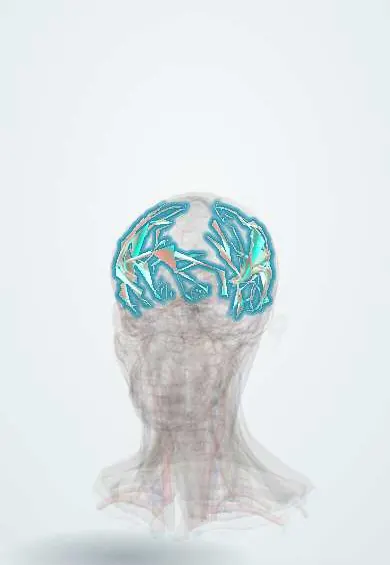
The middle cerebral artery feeds the outer side of the brain, including face, arm and language areas; it is the artery most often blocked in stroke.
Opens in the Durgical app, no sign-up needed.
What it is
The largest branch of the internal carotid, running out in the lateral sulcus over the side of the brain.
What it does
Feeds the face, arm and language areas; the most common site of major strokes.
Key facts
- Origin
- Internal carotid (largest branch)
- Branches
- Lenticulostriate (to basal ganglia, internal capsule); superior and inferior divisions over the lateral surface
- Supplies
- Lateral hemisphere incl. insula, lateral orbital surface and temporal pole: face and arm motor/sensory areas, language (dominant side), optic radiation; deep: putamen, external pallidum, most of the caudate, upper internal capsule
Relations
- Course
- From the end of the internal carotid it runs laterally in the lateral (Sylvian) sulcus (M1), divides near the limen insulae, runs over the insula (M2), turns out between the opercula (M3) and spreads over the lateral surface of the hemisphere (M4)
- Deep
- Insula; its lenticulostriate branches climb through the anterior perforated substance to the basal ganglia and internal capsule
- Superficial
- Frontal, parietal and temporal opercula, which close over it in the lateral sulcus
How to find it
It cannot be felt. M1 runs laterally from the internal carotid under the anterior perforated substance, then the branches climb through the lateral (Sylvian) fissure over the insula; transcranial Doppler finds it through the thin temporal bone above the zygomatic arch, and serial readings are used to watch for vasospasm.
Examination
- Pronator drift: arms held out, palms up, eyes closed; the weak arm drifts down and turns in
- Language: fluency, naming, repetition and comprehension separate Broca from Wernicke aphasia
- Neglect: line bisection, copying a clock face, or extinction when both sides are touched at once
On imaging
Non-contrast CT may show a hyperdense MCA sign (clot in M1) and early loss of the insular ribbon; the ASPECTS score grades early change across the territory; CT angiography looks for a large vessel occlusion.
Common problems
- Ischaemic stroke from a clot: sudden weakness or numbness of the face and arm on one side, trouble speaking.
- Every minute counts: brain cells die quickly without blood.
How it is diagnosed
Emergency CT or MRI and CT angiography to find the blocked artery.
Treatment
- Clot-dissolving medicine within the first hours.
- Removing the clot with a catheter (thrombectomy) for large-artery strokes.
- Rehabilitation and prevention (blood pressure, cholesterol, antiplatelets, quitting smoking).
Clinical notes
- Stroke care is time-critical: imaging decides whether clot-dissolving treatment (thrombolysis) or clot retrieval by catheter (thrombectomy) can help
- Large infarcts can swell over the first days ('malignant' MCA infarction) and cause herniation; decompressive hemicraniectomy is an option
- Homonymous hemianopia appears when the optic radiation in the temporal or parietal lobe is involved
Anatomical variants
- M1 divides into two trunks in most people and into three (trifurcation) in a minority
- An accessory or duplicated middle cerebral artery, arising from the anterior cerebral or internal carotid artery, is uncommon
Memory aid
Stroke recognition: FAST (Face drooping, Arm weakness, Speech difficulty, Time to call for emergency help).
Related conditions
- Ischaemic stroke
- Transient ischaemic attack
- Arterial hypertension
- Vascular dementia
- Brain arteriovenous malformation
- Primary angiitis of the central nervous system
Test yourself
Think of the answer, then tap the question to see it.
What is the typical deficit of a middle cerebral artery stroke?
Weakness and numbness of the opposite face and arm more than the leg, with the eyes turned toward the side of the lesion.
Which aphasias follow strokes of the divisions of the dominant middle cerebral artery?
Superior division: Broca (non-fluent) aphasia. Inferior division: Wernicke (fluent, poor comprehension) aphasia.
Which branches of the middle cerebral artery cause lacunar infarcts and hypertensive bleeds?
The lenticulostriate arteries, small end arteries to the basal ganglia and internal capsule.
What is a malignant middle cerebral artery infarction?
A large infarct that swells over the first days and can cause herniation; decompressive hemicraniectomy is an option.
Educational information, not medical advice. For any symptom, see a health professional.
Its names
- Official Latin (TA2)
- arteria media cerebri TA2 4509
- Official Spanish (DeCS)
- Arteria Cerebral Media DeCS D020768
- English (TA2)
- Middle cerebral artery
- Also called
- sylvian artery
Related
Sources: Terminologia Anatomica (TA2); DeCS; Neuroanatomy, Middle Cerebral Artery; Anatomía humana (4.ª ed.), tomo 1, cap. 26: Vascularización del encéfalo.
Spotted a mistake?We are always improving for the community. Tell us what is wrong and our team will review it.Report a mistake
Representative model, for educational purposes only.