Nervous system · Head and neck
Motor cortex !
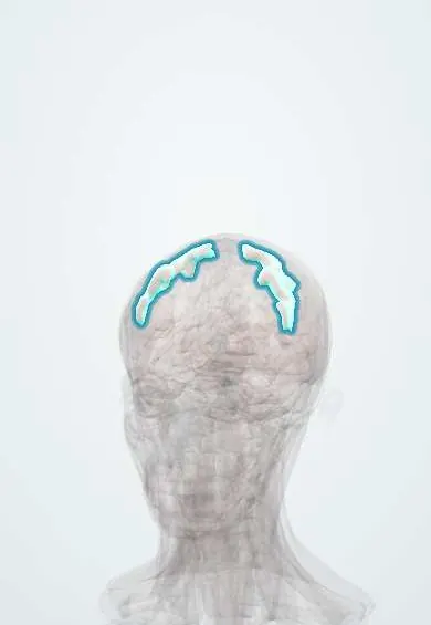
The motor cortex (precentral gyrus) is a strip of brain surface in front of the central groove; it sends commands for voluntary movement to the opposite side.
Official name: Precentral gyrus
Opens in the Durgical app, no sign-up needed.
What it is
The strip of cortex just in front of the central sulcus.
What it does
Sends the commands for voluntary movement to the opposite side of the body.
Key facts
- Where
- Directly in front of the central sulcus
- Blood supply
- MCA (face, arm), ACA (leg)
- Function
- Primary motor cortex (area 4); motor homunculus; controls the opposite side
Relations
- In front
- Precentral sulcus, then the premotor and supplementary motor areas (area 6) and the frontal eye field
- Behind
- Central sulcus, with the sensory cortex (postcentral gyrus) behind it
- Below
- Lateral sulcus; on the dominant side, Broca's area lies just in front of its lower end
- Medial
- Continues over the top onto the medial surface as the front of the paracentral lobule (leg and foot)
- Course
- Its corticospinal fibres descend through the corona radiata, the posterior limb of the internal capsule, the cerebral peduncle, the pons and the medullary pyramids; most cross at the pyramidal decussation
How to find it
On axial MRI the hand area forms a knob shaped like an inverted omega on the precentral gyrus; the superior frontal sulcus ends against the precentral sulcus just in front of it. The precentral gyrus is thicker than the postcentral. On the scalp, the upper end of the central sulcus is estimated about 2 cm behind the midpoint of the nasion–inion line.
Examination
- Pronator drift: arms held out, palms up, eyes closed; one arm drifts down and turns in, a sensitive sign of mild upper motor neuron weakness
- Fine finger movements (finger tapping): slow or clumsy on the side opposite the lesion even when power seems normal
- Babinski sign: the big toe goes up when the sole is stroked, pointing to damage of the corticospinal pathway
On imaging
On axial MRI the central sulcus is found by following the superior frontal sulcus back to the precentral sulcus: the gyrus behind it is precentral, with the hand knob shaped like an inverted omega, and its cortex is thicker than the postcentral. Diffusion MRI shows acute infarcts here (MCA for hand and face, ACA for leg). Before nearby tumour surgery, functional MRI and tractography map it.
Clinical notes
- Jacksonian march: a focal motor seizure spreads along the homunculus (for example hand to arm to face); weakness can follow for hours (Todd paralysis)
- Before a tumour near it is removed, the motor cortex is mapped by direct electrical stimulation or functional MRI
- A small lacunar infarct in the internal capsule can mimic a cortical lesion, but it weakens face, arm and leg equally, without cortical signs such as aphasia or neglect
Memory aid
The homunculus hangs upside down on the side of the brain, with its legs dangling over the top edge into the midline.
Related conditions
- Amyotrophic lateral sclerosis
- Cerebral palsy
- Ischaemic stroke
- Epilepsy and seizures
- Parkinson disease
- Glioblastoma
Test yourself
Think of the answer, then tap the question to see it.
How is the motor homunculus laid out on the precentral gyrus?
Foot and leg on the medial surface (paracentral lobule), then trunk, arm and a large hand, with face, tongue and larynx lowest near the lateral sulcus.
Why does a lesion of the motor cortex spare the forehead?
The upper face is controlled from both hemispheres, so a cortical lesion weakens only the lower face (central facial palsy).
Which artery's stroke weakens the leg most, and which weakens the face and arm more than the leg?
Anterior cerebral artery: leg most; middle cerebral artery: face and arm more than the leg, following the homunculus.
What is a Jacksonian march, and what can follow it?
A focal motor seizure spreading along the homunculus, for example hand to arm to face; weakness lasting hours (Todd paralysis) can follow.
How is the hand area of the motor cortex recognised on axial MRI?
As a knob shaped like an inverted omega on the precentral gyrus, with the superior frontal sulcus ending against the precentral sulcus just in front.
Educational information, not medical advice. For any symptom, see a health professional.
Its names
- Official Latin (TA2)
- gyrus precentralis TA2 5458
- Official Spanish (DeCS)
- Giro precentral DeCS D009044
- English (TA2)
- Precentral gyrus
- Also called
- primary motor cortex
Related
Sources: Terminologia Anatomica (TA2); DeCS; Neuroanatomy, Precentral Gyrus.
Spotted a mistake?We are always improving for the community. Tell us what is wrong and our team will review it.Report a mistake
Representative model, for educational purposes only.