Nervous system · Head and neck
Parietal lobe !
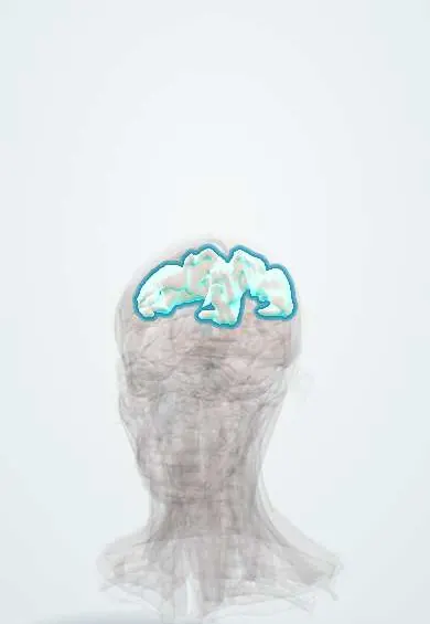
The parietal lobe is the upper middle part of each half of the brain, under the crown of the head; it handles touch, body position, space and attention.
Official name: Superior parietal lobule
Opens in the Durgical app, no sign-up needed.
What it is
The upper middle part of each hemisphere, under the crown of the head.
What it does
Touch, body position, space and attention; the angular gyrus helps with reading and numbers.
Key facts
- Where
- Between the central and parieto-occipital sulci
- Blood supply
- MCA (lateral), ACA (medial)
- Function
- Touch and body position, spatial awareness, reading/calculation (dominant)
Relations
- In front
- Frontal lobe across the central sulcus; the sensory cortex (postcentral gyrus) is its first gyrus
- Behind
- Occipital lobe: the parieto-occipital sulcus on the medial surface; on the lateral surface only an imaginary line to the preoccipital notch
- Below
- Temporal lobe across the back of the lateral sulcus; the supramarginal gyrus caps the end of the lateral sulcus and the angular gyrus the end of the superior temporal sulcus
- Medial
- Precuneus and the back of the paracentral lobule, facing the falx cerebri
- Superficial
- Parietal bone, under the crown of the head
- Deep
- Body and atrium of the lateral ventricles, with the upper optic radiation in the white matter beside them
How to find it
The intraparietal sulcus runs back from the postcentral sulcus and splits the lobe into the superior parietal lobule above and the inferior parietal lobule (supramarginal and angular gyri) below. On axial MRI, the sensory cortex behind the hand knob and the bracket sign mark its front edge; the parieto-occipital sulcus is clearest on sagittal images.
Examination
- Double simultaneous stimulation: touches or finger movements on both sides at once are reported only on one side (extinction), an early sign of neglect
- Line bisection, cancellation and clock drawing: the mark is shifted, or items are missed, on the side opposite the lesion
- Stereognosis (naming a coin or key by touch, eyes closed) and graphesthesia (numbers traced on the palm): failure with intact touch suggests a parietal lesion
On imaging
MRI is preferred; CT comes first in stroke and head injury. Landmarks: the postcentral gyrus behind the central sulcus, the intraparietal sulcus, and the parieto-occipital sulcus on the medial surface (best on sagittal images). Typical findings: middle cerebral artery infarcts of the inferior parietal lobule, parietal and precuneus atrophy in Alzheimer disease, parieto-occipital oedema in PRES.
Common problems
- Head injury and tumours here change personality, judgement and planning.
- Front-lobe dementia changes behaviour first.
How it is diagnosed
Neurological and memory tests; CT or MRI.
Treatment
- Surgery for tumours when possible.
- Rehabilitation and support.
Clinical notes
- Neglect is more frequent and lasting after right than left lesions, as the right hemisphere attends to both sides of space; it slows rehabilitation and raises the risk of falls
- Left inferior parietal lesions cause ideomotor apraxia: trouble miming the use of objects on command despite normal strength
- Bilateral parieto-occipital damage, for example border-zone infarcts after a drop in blood pressure, gives Balint syndrome: simultanagnosia, optic ataxia and oculomotor apraxia
Related conditions
- MELAS (mitochondrial encephalomyopathy, lactic acidosis and stroke-like episodes)
- Neurocysticercosis
- Alzheimer disease
- Functional neurological symptom disorder (conversion disorder)
Test yourself
Think of the answer, then tap the question to see it.
What does a non-dominant (usually right) parietal lobe lesion cause?
Neglect of the opposite side of space and body, often with denial of the deficit (anosognosia) and dressing or constructional apraxia.
What are the four features of Gerstmann syndrome, and where is the lesion?
Agraphia, acalculia, finger agnosia and left–right confusion, from a lesion of the dominant (usually left) angular gyrus.
Which visual field defect does damage deep in the parietal lobe give?
A contralateral inferior quadrantanopia («pie on the floor»), from the upper fibres of the optic radiation.
What does failure of stereognosis and graphesthesia with intact basic sensation suggest?
A lesion of the parietal cortex behind the sensory cortex.
What is extinction on double simultaneous stimulation?
Touches on both sides at once are reported only on one side, an early sign of neglect from a parietal lesion.
Educational information, not medical advice. For any symptom, see a health professional.
Its names
- Official Latin (TA2)
- lobulus parietalis superior TA2 5476
- Official Spanish (DeCS)
- Lóbulo Parietal DeCS D010296
- English (TA2)
- Superior parietal lobule
Related
Sources: Terminologia Anatomica (TA2); DeCS; Neuroanatomy, Cerebral Cortex; Neuroanatomy, Cerebral Hemisphere.
Spotted a mistake?We are always improving for the community. Tell us what is wrong and our team will review it.Report a mistake
Representative model, for educational purposes only.