Skeletal system · Head and neck
Frontal sinus !
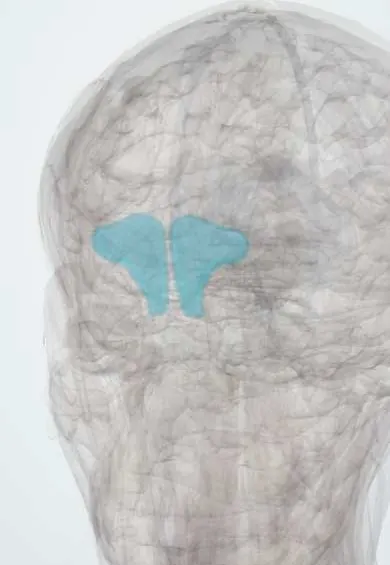
The frontal sinus is an air space in the frontal bone above the eyes; it lightens the skull and can fill up and hurt in sinusitis.
Official name: Sinus of frontal bone
Opens in the Durgical app, no sign-up needed.
What it is
An air space in the frontal bone above the eyes.
What it does
Lightens the skull; it can fill and hurt in sinusitis.
Key facts
- Where
- Frontal bone above the orbits; drains to the middle meatus
- Blood supply
- Supraorbital and anterior ethmoidal arteries
- Nerve
- V1 (supraorbital)
- Function
- Air space; absent at birth, develops ~7 years
Relations
- Behind
- Anterior cranial fossa: dura mater and the frontal lobes
- Below
- Roofs of the orbits and, medially, the frontal recess opening into the anterior ethmoid cells and middle meatus
- In front
- Anterior table of the frontal bone, the brow and the supraorbital and supratrochlear nerves
- Medial
- The other frontal sinus across a thin septum, usually off the midline
How to find it
It lies behind the brow, above the medial part of each orbit; tenderness over the forehead or the sinus floor at the upper inner corner of the orbit can accompany frontal sinusitis.
Examination
- Pressure or tapping on the forehead and on the sinus floor at the upper inner corner of the orbit: tenderness on one side supports frontal sinusitis, but the sign is not specific and imaging or endoscopy settles it
On imaging
CT shows its size, septa, fluid and the frontal recess; MRI with contrast is used when spread into the orbit or the cranial cavity is suspected.
Common problems
- Sinusitis: blocked nose, thick discharge, pressure in the cheeks or teeth.
- Most acute sinusitis is viral; some becomes chronic.
How it is diagnosed
Symptoms and exam; nasal endoscopy and CT for chronic or complicated cases.
Treatment
- Saline rinses, nasal steroid sprays, pain relief; antibiotics only for bacterial cases.
- Endoscopic sinus surgery for chronic sinusitis.
Clinical notes
- A mucocele forms when its outflow is blocked (often after trauma or surgery) and can expand into the orbit, pushing the eye down and out
- Frontal sinus fractures involving the posterior table or the outflow tract carry a risk of CSF leak, meningitis and later mucocele
- Endoscopic frontal sinus surgery works through the frontal recess, whose shape depends on the agger nasi and other ethmoid cells around it
Anatomical variants
- Absent on one side in a minority of people and on both sides in fewer; figures vary between studies
- Size and septa vary widely, and the two sides are usually unequal
Development
It grows up from the anterior ethmoid region into the frontal bone, which is why it drains through the ethmoid area; it keeps enlarging until after puberty, so Pott puffy tumour is seen mostly in adolescents.
Related conditions
Test yourself
Think of the answer, then tap the question to see it.
What is Pott puffy tumour in frontal sinusitis?
Spread of frontal sinusitis into the frontal bone, causing osteomyelitis with a subperiosteal abscess (soft forehead swelling), often with intracranial spread.
How can infection pass from the frontal sinus to the brain?
Through valveless veins across the posterior wall, causing meningitis, epidural or subdural empyema or a frontal lobe abscess.
Where does the frontal sinus drain, and why can ethmoid disease block it?
Through the frontal recess into the middle meatus, a path bordered by anterior ethmoid cells.
What can a blocked frontal sinus outflow form, and where can it expand?
A mucocele, which can expand into the orbit and push the eye down and out.
Educational information, not medical advice. For any symptom, see a health professional.
Its names
- Official Latin (TA2)
- sinus ossis frontalis TA2 549
- Official Spanish (DeCS)
- Seno Frontal DeCS D005626
- English (TA2)
- Sinus of frontal bone
- Also called
- bony frontal sinus · sinus frontalis
Related
Sources: Terminologia Anatomica (TA2); DeCS; Anatomy, Head and Neck, Nose Paranasal Sinuses; Anatomy, Head and Neck, Nose Sinuses.
Spotted a mistake?We are always improving for the community. Tell us what is wrong and our team will review it.Report a mistake
Representative model, for educational purposes only.