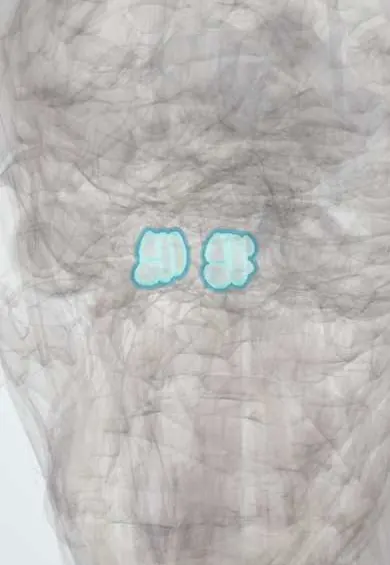
Skeletal system · Head and neck
Ethmoid cells !
The ethmoid cells are many small air spaces between the eyes; they drain into the nose, and an infection there can spread to the eye socket.
Official name: Anterior cells of ethmoid bone
Opens in the Durgical app, no sign-up needed.
What it is
Many small air cells between the eyes.
What it does
Drain into the nose; infection can spread to the eye socket.
Key facts
- Where
- Between the orbits (anterior, middle, posterior groups)
- Blood supply
- Ethmoidal arteries
- Nerve
- V1 (nasociliary), V2
- Function
- Air cells; thin orbital wall (lamina papyracea) lets infection reach the orbit
Relations
- Lateral
- Orbit through the lamina papyracea; the medial rectus, and the anterior and posterior ethmoidal arteries crossing at the roof
- Above
- Ethmoid roof (fovea ethmoidalis) and cribriform plate, separating them from the anterior cranial fossa and frontal lobes
- Medial
- Superior and middle conchae and the upper nasal cavity
- Behind
- Sphenoid sinus; posterior cells can lie next to the optic nerve
- Below
- Maxillary sinus and the ostiomeatal complex in the middle meatus
- In front
- Frontal recess and lacrimal bone
How to find it
They cannot be felt; tenderness at the inner corner of the eye can accompany ethmoid sinusitis. Endoscopically the cells are opened through the middle meatus, usually starting at the ethmoid bulla.
Examination
- Eye movements, vision, colour vision and pupils: pain on eye movement, restricted movement, proptosis or a relative afferent pupillary defect mark orbital rather than preseptal cellulitis
On imaging
Contrast CT of the orbits and sinuses separates preseptal from orbital cellulitis and shows a subperiosteal abscess along the lamina papyracea; coronal CT maps the cells before surgery.
Common problems
- Sinusitis: blocked nose, thick discharge, pressure in the cheeks or teeth.
- Most acute sinusitis is viral; some becomes chronic.
How it is diagnosed
Symptoms and exam; nasal endoscopy and CT for chronic or complicated cases.
Treatment
- Saline rinses, nasal steroid sprays, pain relief; antibiotics only for bacterial cases.
- Endoscopic sinus surgery for chronic sinusitis.
Clinical notes
- In endoscopic sinus surgery the anterior ethmoidal artery at the skull base and the lamina papyracea are key landmarks; injury can cause an orbital haematoma or CSF leak
- The ostiomeatal complex is the shared drainage path of the frontal, maxillary and anterior ethmoid sinuses, so a blockage there can affect all three
Anatomical variants
- Agger nasi cell: the most anterior ethmoid cell, in front of the middle concha attachment, present in most people
- Haller (infraorbital ethmoid) cells along the orbital floor can narrow the outflow of the maxillary sinus
- Onodi (sphenoethmoidal) cells extend above or beside the sphenoid sinus and can lie against the optic nerve, a surgical risk
- Concha bullosa: an air-filled middle concha, which can narrow the middle meatus
Development
Present at birth and enlarging through childhood, which is why ethmoid sinusitis is seen even in infants.
Related conditions
Test yourself
Think of the answer, then tap the question to see it.
What is the most common source of orbital cellulitis in children, and how does it spread?
Ethmoid sinusitis, spreading through the thin lamina papyracea and its veins.
What are the Chandler stages of orbital spread from the ethmoid cells?
Preseptal cellulitis, orbital cellulitis, subperiosteal abscess, orbital abscess, cavernous sinus thrombosis.
Where do the anterior, middle and posterior ethmoid cells drain?
Anterior and middle cells to the middle meatus (ostiomeatal complex); posterior cells to the superior meatus.
Why is an Onodi cell a surgical risk among the ethmoid cells?
This posterior ethmoid cell extends above or beside the sphenoid sinus and can lie against the optic nerve.
Educational information, not medical advice. For any symptom, see a health professional.
Its names
- Official Latin (TA2)
- cellulae anteriores ossis ethmoidei TA2 729
- Official Spanish (DeCS)
- Senos Etmoidales DeCS D005005
- English (TA2)
- Anterior cells of ethmoid bone
- Also called
- ethmoid sinus · bony anterior ethmoidal cells · sinus ethmoidei anteriores · cellulae ethmoidales anteriores osseae
Sources: Terminologia Anatomica (TA2); DeCS; Anatomy, Head and Neck, Nose Paranasal Sinuses; Anatomy, Head and Neck, Nose Sinuses.
Spotted a mistake?We are always improving for the community. Tell us what is wrong and our team will review it.Report a mistake
Representative model, for educational purposes only.