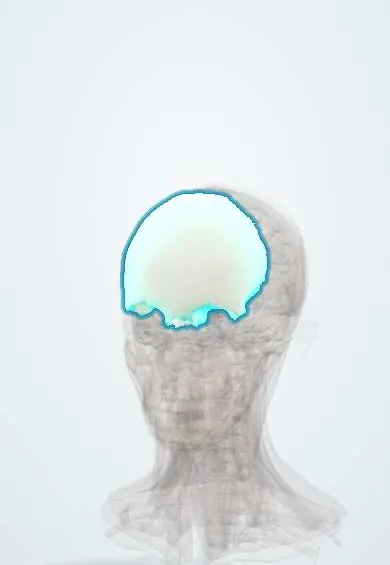
Skeletal system · Head and neck
Frontal bone !
The frontal bone is the forehead bone; it forms the front of the skull and the roof of the eye sockets and protects the front of the brain.
Opens in the Durgical app, no sign-up needed.
What it is
The forehead bone; it forms the front of the skull and the roof of the eye sockets.
What it does
Protects the frontal lobes and holds the frontal sinus.
Key facts
- Joints with
- Parietals, sphenoid, ethmoid, nasals, maxillae, zygomatics, lacrimals
- Landmarks
- Glabella, supraorbital margins and notch, frontal sinus, orbital plates; frontal crest (falx cerebri); foramen cecum; fossa for the lacrimal gland; trochlear fovea (superior oblique pulley)
Relations
- Deep
- Frontal lobes, covered by dura mater; the superior sagittal sinus runs in the midline groove on its inner surface, and the falx cerebri attaches to the frontal crest
- Below
- The orbits and their contents beneath the thin orbital plates; the ethmoid bone fits into the notch between the plates, and the nasal bones meet it at the nasion
- Behind
- Parietal bones at the coronal suture; laterally the greater wing of the sphenoid at the pterion
- Superficial
- Frontalis and the skin of the forehead; the supraorbital and supratrochlear nerves and vessels run up from the orbital rim
How to find it
The forehead, the brow ridges and the supraorbital margins are felt directly. The glabella is the smooth area between the eyebrows; the supraorbital notch is pressed on the medial part of the upper orbital rim.
Examination
- Supraorbital pressure: firm pressure over the notch is a central painful stimulus used when scoring the motor response of the Glasgow Coma Scale; a hand reaching up toward it is a localising response
On imaging
CT with bone windows and 3D reconstructions shows frontal sinus fractures and whether the posterior table and the sinus outflow tract are involved; skull radiographs show the sinus outline but miss many fractures.
Clinical notes
- Pott puffy tumour: soft forehead swelling from a subperiosteal abscess with osteomyelitis of the frontal bone, a complication of frontal sinusitis, mostly in adolescents
- Injury to the frontal sinus outflow tract in a fracture predisposes to a later mucocele
- Premature fusion of the metopic suture gives a narrow, triangular forehead (trigonocephaly)
- The supraorbital nerve can be blocked at the notch to numb the forehead and front of the scalp for procedures
Anatomical variants
- A metopic suture that stays open into adulthood (a small minority of adults); on radiographs it can be mistaken for a fracture
- A supraorbital foramen instead of a notch, on one or both sides
Development
Ossifies in membrane from two primary centres, one above each orbit, joined by the metopic suture, which normally closes in infancy (texts differ on the exact age). The anterior fontanelle usually closes by about 18 to 24 months.
Related conditions
Test yourself
Think of the answer, then tap the question to see it.
Which fracture of the frontal sinus walls risks a CSF leak and meningitis, and why?
A fracture of the posterior table: it lies against the dura, so it can tear it. An anterior table fracture mainly changes the forehead contour.
What is Pott puffy tumour of the frontal bone?
A soft forehead swelling from a subperiosteal abscess with osteomyelitis of the frontal bone, a complication of frontal sinusitis, mostly in adolescents.
Which nerve leaves the orbit through the supraorbital notch of the frontal bone, and how is pressure there used clinically?
The supraorbital nerve (V1). Firm supraorbital pressure is a central painful stimulus for the motor score of the Glasgow Coma Scale.
What head shape follows premature fusion of the metopic suture of the frontal bone?
Trigonocephaly: a narrow, triangular forehead. At birth the frontal bone is two halves joined by the metopic suture.
Educational information, not medical advice. For any symptom, see a health professional.
Its names
- Official Latin (TA2)
- os frontale TA2 520
- Official Spanish (DeCS)
- Hueso Frontal DeCS D005624
- English (TA2)
- Frontal bone
Related
Sources: Terminologia Anatomica (TA2); DeCS; Anatomy, Head and Neck: Frontal Bone; Anatomy, Head and Neck, Skull.
Spotted a mistake?We are always improving for the community. Tell us what is wrong and our team will review it.Report a mistake
Representative model, for educational purposes only.