Nervous system · Head and neck
Frontal lobe !
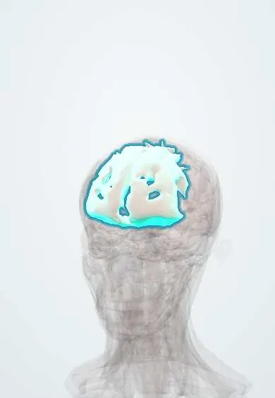
The frontal lobe is the front part of each half of the brain, behind the forehead; it handles planning, decisions, behaviour, attention and movement.
Official name: Superior frontal gyrus
Opens in the Durgical app, no sign-up needed.
What it is
The front part of each cerebral hemisphere, behind the forehead.
What it does
Planning, decisions, behaviour, attention and (with the motor strip) movement.
Key facts
- Where
- In front of the central sulcus
- Blood supply
- ACA (medial), MCA (lateral)
- Function
- Movement, planning, judgement, personality, speech production (Broca)
Relations
- In front
- Frontal bone, with the frontal sinus in front of its lower part
- Behind
- Parietal lobe across the central sulcus; the motor cortex (precentral gyrus) is its last gyrus before the sulcus
- Below
- Orbital surface on the roof of the orbit (anterior cranial fossa), with the olfactory bulb and tract in the olfactory sulcus; the gyrus rectus lies medial to this sulcus and the orbital gyri lateral to it, around an H-shaped orbital sulcus; further back, the temporal lobe across the lateral sulcus
- Medial
- Falx cerebri in the longitudinal fissure; on the medial surface, the cingulate gyrus with the corpus callosum below it
- Deep
- Frontal horn of the lateral ventricles, head of the caudate nucleus and anterior limb of the internal capsule
How to find it
It runs from the frontal pole back to the central sulcus and down to the lateral sulcus, with the superior, middle and inferior frontal gyri in front of the precentral gyrus. On axial MRI the central sulcus is found where the superior frontal sulcus meets the precentral sulcus, behind the hand knob (an inverted omega), and one gyrus in front of the bracket sign of the cingulate sulcus.
Examination
- Frontal release signs (grasp, palmomental, snout, rooting): infant reflexes that reappear with frontal or diffuse cortical disease; an isolated palmomental reflex can be normal in older adults
- Luria fist–edge–palm sequence and go/no-go task: failure to learn or hold the sequence, or to inhibit a response, points to executive (prefrontal) dysfunction
- Letter fluency (words starting with one letter in a minute): a low score suggests frontal, especially left frontal, dysfunction
On imaging
MRI is preferred; CT comes first in head injury and acute stroke. Typical findings: contusions on the orbital surface after a blow to the head, frontal-predominant atrophy in frontotemporal dementia, an olfactory groove meningioma pressing from below, and infarcts of the anterior cerebral artery territory on the medial surface, shown on diffusion-weighted MRI.
Common problems
- Head injury and tumours here change personality, judgement and planning.
- Front-lobe dementia changes behaviour first.
How it is diagnosed
Neurological and memory tests; CT or MRI.
Treatment
- Surgery for tumours when possible.
- Rehabilitation and support.
Clinical notes
- Frontal lobe seizures are brief, often arise from sleep, with bizarre thrashing or posturing and quick recovery; they are easily mistaken for psychogenic events
- Olfactory groove meningiomas can grow large under the frontal lobes before diagnosis, presenting with personality change and loss of smell
- Contusions favour the orbital frontal surface and the temporal poles, where the brain slides over the rough floor of the anterior and middle cranial fossae
Development
Grows from the telencephalon, the front part of the forebrain vesicle. The prefrontal cortex is among the last areas to mature: its myelination and synaptic pruning go on into the early twenties.
Related conditions
- Smith–Lemli–Opitz syndrome
- Sanfilippo syndrome (mucopolysaccharidosis type III)
- Fragile X syndrome
- GM1 gangliosidosis
- Prion diseases (Creutzfeldt–Jakob)
- Traumatic brain injury
- Epilepsy and seizures
- Neurocysticercosis
- Glioblastoma
- Acute alcohol (ethanol) intoxication
- Benzodiazepine overdose
- Dementia (major neurocognitive disorder)
- Toxoplasmosis
- Brain abscess
- Free-living amoeba infections (Naegleria meningoencephalitis, Acanthamoeba)
- Status epilepticus
- Tuberous sclerosis complex
- Frontotemporal dementia
- Fetal alcohol spectrum disorder
- Infantile spasms (West syndrome)
- Rett syndrome
- Major depressive disorder
- Suicidal behaviour and suicide risk
- Bipolar disorder
- Schizophrenia
- Generalized anxiety disorder
- Alcohol use disorder
- Alcohol withdrawal and delirium tremens
- Opioid use disorder, overdose and withdrawal
- Post-traumatic stress disorder (PTSD) and acute stress disorder
- Delirium
- Obsessive-compulsive disorder (OCD)
- Attention-deficit/hyperactivity disorder (ADHD)
- Personality disorders (clusters A, B and C)
- Persistent depressive disorder (dysthymia)
- Schizoaffective, schizophreniform, brief psychotic and delusional disorders
- Adjustment disorder
- Functional neurological symptom disorder (conversion disorder)
- Factitious disorder and malingering
- Stimulant use disorder (cocaine, amphetamines)
- Cannabis use disorder
- Tobacco use disorder
- Sedative-hypnotic (benzodiazepine) use disorder and withdrawal
- Catatonia
- Conduct disorder and oppositional defiant disorder
- Intimate partner and sexual violence (mental-health care)
- Disruptive mood dysregulation disorder (DMDD)
- Dissociative disorders
- Hallucinogen and dissociative drug intoxication (LSD, PCP, ketamine)
- Gambling disorder
- Paraphilic disorders
Test yourself
Think of the answer, then tap the question to see it.
What does damage to the frontal lobe change more than strength?
Personality, judgement and planning: apathy or disinhibition, poor insight and impulsive choices; Phineas Gage is the classic historical case.
Which way do the eyes turn with an acute destructive lesion of the frontal eye field, and with a seizure there?
Toward the lesion with a destructive lesion; away from it with a seizure, as the frontal eye field (area 8) drives gaze to the opposite side.
What triad does a medial frontal lobe lesion in anterior cerebral artery territory give?
Leg-predominant weakness, apathy and urinary incontinence, as cortical bladder control lies on the medial surface.
What are frontal release signs?
Infant reflexes (grasp, palmomental, snout, rooting) that reappear with frontal or diffuse cortical disease; an isolated palmomental reflex can be normal in older adults.
Which tumour can grow large under the frontal lobes, presenting with personality change and loss of smell?
Olfactory groove meningioma, beneath the orbital surface of the frontal lobe where the olfactory bulb and tract lie.
Educational information, not medical advice. For any symptom, see a health professional.
Its names
- Official Latin (TA2)
- gyrus frontalis superior TA2 5456
- Official Spanish (DeCS)
- Lóbulo Frontal DeCS D005625
- English (TA2)
- Superior frontal gyrus
Related
Sources: Terminologia Anatomica (TA2); DeCS; Neuroanatomy, Frontal Cortex; Neuroanatomy, Cerebral Cortex; 13.1 The Embryologic Perspective (OpenStax (Rice University), 2022) · adapted, CC BY 4.0; Tratado de anatomía humana (9.ª ed.), tomo II, Libro V, cap. VI: Cerebro (configuración exterior e interior, ventrículos, formaciones coroideas, epífisis).
Spotted a mistake?We are always improving for the community. Tell us what is wrong and our team will review it.Report a mistake
Representative model, for educational purposes only.