Nervous system · Head and neck
Dura mater !
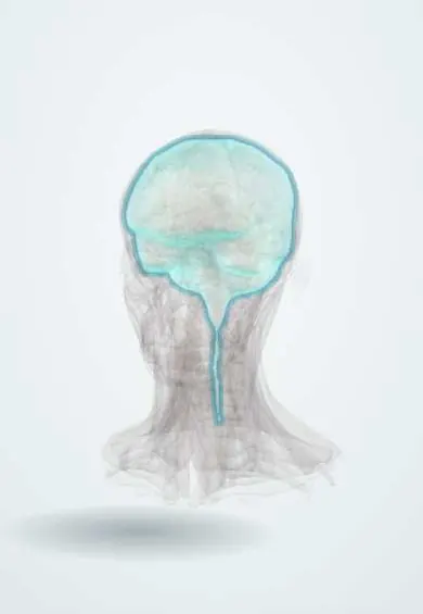
The dura mater is the tough outer membrane lining the inside of the skull; it protects the brain, and bleeding above or below it can press on the brain.
Official name: Cranial dura
Opens in the Durgical app, no sign-up needed.
What it is
The tough outer membrane lining the inside of the skull.
What it does
Protects the brain; bleeding outside it (epidural) or under it (subdural) presses on the brain.
Key facts
- Where
- Lines the inside of the skull; folds: falx, tentorium
- Blood supply
- Middle meningeal artery (main)
- Nerve
- V1–V3 (supratentorial), C2–C3 and X (posterior fossa): source of headache
- Function
- Protects the brain; holds the venous sinuses
Relations
- Superficial
- Inner surface of the skull; the epidural space is only a potential space, opened by blood or pus
- Deep
- Arachnoid, across a potential subdural space crossed by bridging veins; beneath it the subarachnoid space holds CSF
- Contents
- Between its layers: the dural venous sinuses; in its outer layer: the meningeal arteries, chiefly the middle meningeal artery, and meningeal nerves
- Below
- At the foramen magnum the inner layer continues as the spinal dura, which, unlike the cranial dura, has a true epidural space with fat and veins
How to find it
In a craniotomy it is the tough, whitish layer directly under the bone, with the middle meningeal vessels on its outer surface. The pterion, over the frontal branch of the middle meningeal artery, lies about two finger-breadths above the zygomatic arch and a thumb's breadth behind the frontal process of the zygomatic bone.
On imaging
On CT, epidural blood is biconvex and limited by sutures; subdural blood is crescentic and may cross sutures; acute blood is bright and a chronic subdural becomes dark. On contrast MRI, smooth diffuse enhancement suggests low CSF pressure; thick or nodular enhancement suggests tumour or inflammation.
Common problems
- Epidural haematoma: bleeding between skull and dura, usually from the middle meningeal artery after a blow; the person may seem fine at first, then worsens.
- Subdural haematoma: bleeding under the dura from torn veins, common in older adults and people on blood thinners.
How it is diagnosed
Emergency CT of the head.
Treatment
- Observation for small bleeds.
- Urgent craniotomy or burr holes to remove the blood.
Clinical notes
- Meningioma: a dura-based tumour of arachnoid cells over the convexity, falx or skull base; contrast MRI often shows a dural tail
- Post-dural-puncture headache: CSF leaks through a hole in the spinal dura after lumbar puncture or spinal anaesthesia; worse upright, easier lying down
- Referred pain: dura above the tentorium refers pain to the forehead and face (trigeminal), dura of the posterior fossa to the back of the head and neck
- Dural tears in skull base fractures cause CSF leak from the nose or ear and a risk of meningitis
Related conditions
Test yourself
Think of the answer, then tap the question to see it.
What are the two layers of the dura mater, and what lies between them?
An outer periosteal and an inner meningeal layer; they part to enclose the dural venous sinuses, and the inner layer folds in as the falx and tentorium.
What is the source and CT shape of an epidural haematoma?
Usually the middle meningeal artery at the pterion; lens-shaped (biconvex) and stopped by the sutures, where the dura is firmly attached.
What is the source and CT shape of a subdural haematoma?
Torn bridging veins; crescent-shaped, crossing sutures but not the falx; common in older adults, as brain atrophy stretches the veins.
Which nerves make the dura mater pain-sensitive?
V1–V3 above the tentorium; C2–C3 and the vagus in the posterior fossa; the dura is a main source of headache, while brain tissue is not.
What causes post-dural-puncture headache, and how does posture change it?
CSF leaks through a hole in the spinal dura after lumbar puncture or spinal anaesthesia; it is worse upright and easier lying down.
Educational information, not medical advice. For any symptom, see a health professional.
Its names
- Official Latin (TA2)
- dura cranialis TA2 5371
- Official Spanish (DeCS)
- Duramadre DeCS D004388
- English (TA2)
- Cranial dura
- Also called
- cranial dura mater · dura mater encephali · dura encephali
Related
Sources: Terminologia Anatomica (TA2); DeCS; Anatomy, Head and Neck, Dura Mater; Neuroanatomy, Cranial Meninges.
Spotted a mistake?We are always improving for the community. Tell us what is wrong and our team will review it.Report a mistake
Representative model, for educational purposes only.