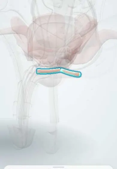
Superficial transverse perineal !
The superficial transverse perineal is a thin muscle crossing the perineum to the perineal body; it steadies the perineal body and can tear in childbirth.
Official name: Superficial transverse perineal muscle
Opens in the Durgical app, no sign-up needed.
What it is
A narrow muscle running across from the sitting bone to the perineal body.
What it does
Steadies the perineal body.
Key facts
- Origin
- Ischial tuberosity (inner surface)
- Insertion
- Perineal body
- Action
- Steadies the perineal body
- Nerve
- Perineal branch of pudendal nerve (S2–S4)
- Blood supply
- Perineal branch of internal pudendal artery
Relations
- Lateral
- The ischial tuberosity
- Medial
- The perineal body
- In front
- The ischiocavernosus and the bulbospongiosus, with the perineal membrane between them
- Behind
- The ischioanal fossa and the external anal sphincter
- Deep
- The posterior edge of the perineal membrane and the deep transverse perineal muscle
How to find it
Not felt on its own. Its line is the imaginary transverse line just in front of the ischial tuberosities, through the central point of the perineum. In dissection, once Colles fascia is lifted, it is the slender strip running from the inner side of the tuberosity to the perineal body along the back edge of the perineal membrane.
On imaging
Too thin to see reliably on imaging. It runs along the line between the ischial tuberosities, which radiographs and CT show as its landmarks. After a second-degree tear it is assessed with the perineal body: transperineal or endoanal ultrasound checks the perineal body and the anal sphincter behind it. MRI images the perineum when a fistula, abscess or mass is suspected.
Common problems
- Straddle injuries (falling astride a bar or a beam) can bruise the perineum and tear the urethra.
- Perineal pain or numbness, for example from long cycling.
How it is diagnosed
Examination; ultrasound or MRI when needed.
Treatment
- Depends on the cause: rest, a better bicycle seat, or treatment of a urethral injury by a urologist.
Clinical notes
- Colles fascia fuses with the back edge of the perineal membrane along this muscle, so blood or urine in the superficial perineal pouch does not spread back into the anal triangle
- Its line is the boundary used to describe perineal regions and to plan perineal incisions
Anatomical variants
- Often thin and poorly developed, and it may be absent on one or both sides
Related conditions
Test yourself
Think of the answer, then tap the question to see it.
Along which line does the superficial transverse perineal muscle run, and what does it divide?
The line between the ischial tuberosities; it divides the perineum into the urogenital triangle in front and the anal triangle behind.
Why does blood or urine in the superficial perineal pouch not spread back into the anal triangle?
Colles fascia fuses with the back edge of the perineal membrane along the superficial transverse perineal muscle.
In which perineal tear is the superficial transverse perineal muscle torn?
Second-degree tears, together with the perineal body, and it is repaired with it.
Educational information, not medical advice. For any symptom, see a health professional.
Its names
- Official Latin (TA2)
- musculus transversus superficialis perinei TA2 2416
- Official Spanish (DeCS)
- Músculo transverso superficial del periné
- English (TA2)
- Superficial transverse perineal muscle
- Also called
- Theile's muscle
Related
Sources: Terminologia Anatomica (TA2); Anatomy, Abdomen and Pelvis: Superficial Perineal Space; Anatomy of the Human Body (20th ed.): Surface Markings of the Perineum (Lea & Febiger (Gray's Anatomy, 1918), 1918) · adapted, Public domain.
Spotted a mistake?We are always improving for the community. Tell us what is wrong and our team will review it.Report a mistake
Representative model, for educational purposes only.