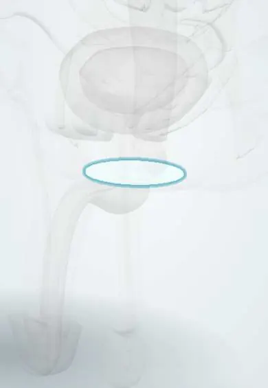
Perineal membrane !
The perineal membrane is a tough sheet stretched across the front of the pelvic outlet; it supports the urethra and the erectile tissue of the vulva.
Opens in the Durgical app, no sign-up needed.
What it is
A tough triangular sheet stretched between the ischiopubic rami, pierced by the urethra.
What it does
Anchors the root of the penis (bulb and crura) from below; the urethra crosses it as the membranous urethra.
Key facts
- Where
- Triangular fibrous sheet across the anterior pelvic outlet, between the ischiopubic rami
- Attaches
- Ischiopubic rami laterally; perineal body posteriorly; pierced by the urethra and the vagina
- Function
- Separates the superficial and deep perineal spaces; attaches the vestibular bulbs and clitoral crura; supports the distal urethra and vagina
Relations
- Above
- The deep perineal pouch with the external urethral sphincter, then the levator ani
- Below
- The superficial perineal pouch with the erectile tissue, its muscles and the Bartholin glands
- Lateral
- The ischiopubic rami, to which it is attached
- Behind
- Its free back edge, fixed to the perineal body, where the transverse perineal muscles lie
- In front
- A thickened front edge (transverse perineal ligament) leaving a gap below the pubic symphysis for the deep dorsal vein of the clitoris
How to find it
In dissection it appears once the superficial pouch is cleared: a tough triangular sheet across the front of the pelvic outlet, attached to both ischiopubic rami, with the perineal body at the middle of its back edge.
On imaging
Its position decides how a male urethral injury is classed: a retrograde urethrogram shows contrast leaking below it (bulbar urethra) or above it (membranous and prostatic urethra). Pelvic floor MRI can show it as a thin low-signal sheet.
Common problems
- Straddle injuries (falling astride a bar or a beam) can bruise the perineum and tear the urethra.
- Perineal pain or numbness, for example from long cycling.
How it is diagnosed
Examination; ultrasound or MRI when needed.
Treatment
- Depends on the cause: rest, a better bicycle seat, or treatment of a urethral injury by a urologist.
Clinical notes
- Male: a straddle injury tears the bulbar urethra below the membrane; urine then fills the superficial pouch and spreads to the scrotum, penis and lower abdominal wall, not the thighs
- Male: a pelvic fracture tears the urethra above the membrane; urine then leaks into the pelvis around the bladder and prostate
- Female: with the perineal body and levator ani it forms the lowest level of vaginal support (DeLancey level III), holding the distal vagina and urethra in place
Related conditions
Test yourself
Think of the answer, then tap the question to see it.
Which structures lie above and which below the perineal membrane in the female?
Above (deep pouch): external urethral sphincter, compressor urethrae, deep transverse perineal. Below (superficial pouch): clitoral crura, vestibular bulbs, Bartholin glands, their muscles.
In a male straddle injury, where is the urethra torn and where does urine spread?
The bulbar urethra below the perineal membrane; urine fills the superficial pouch and spreads to the scrotum, penis and lower abdominal wall, not the thighs.
In a male pelvic fracture, where is the urethra torn relative to the perineal membrane?
Above the perineal membrane; urine leaks into the pelvis around the bladder and prostate.
Which level of vaginal support does the perineal membrane form?
DeLancey level III, with the perineal body and levator ani, holding the distal vagina and urethra.
Educational information, not medical advice. For any symptom, see a health professional.
Its names
- Official Latin (TA2)
- membrana perinei TA2 2444
- Official Spanish (DeCS)
- Membrana perineal
- English (TA2)
- Perineal membrane
- Also called
- fascia inferior diaphragmatis urogenitalis · fascia externa diaphragmatis urogenitalis
Related
Sources: Terminologia Anatomica (TA2); Anatomy, Abdomen and Pelvis: Deep Perineal Space; 11.4 Axial Muscles of the Abdominal Wall, and Thorax (OpenStax (Rice University), 2022) · adapted, CC BY 4.0.
Spotted a mistake?We are always improving for the community. Tell us what is wrong and our team will review it.Report a mistake
Representative model, for educational purposes only.