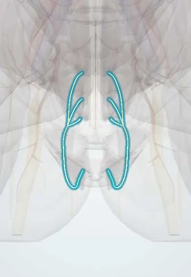
Pudendal nerve !
The pudendal nerve comes from S2-S4 and curves into the perineum; it supplies the pelvic floor, the sphincters and genital and anal feeling.
Opens in the Durgical app, no sign-up needed.
What it is
A nerve from S2-S4 that leaves the pelvis and curves into the perineum.
What it does
Supplies the pelvic floor, the sphincters and feeling in the genital and anal area.
Key facts
- Roots
- Ventral rami of S2–S4; leaves pelvis via greater sciatic foramen, enters perineum via lesser sciatic foramen, in the pudendal (Alcock) canal
- Moves
- External anal sphincter, external urethral sphincter, perineal muscles, part of levator ani
- Feeling from
- Penis or clitoris (dorsal nerve), perineum, posterior scrotum or labia, anal canal below the pectinate line
Relations
- Course
- Leaves through the greater sciatic foramen below the piriformis, crosses behind the sacrospinous ligament at the ischial spine, enters the perineum and runs in the pudendal canal, giving the inferior rectal nerve, then the perineal nerve and the dorsal nerve of the penis or clitoris
- Behind
- At the ischial spine, the sacrotuberous ligament; the nerve lies between the two ligaments, a known site of entrapment
- Lateral
- In the pudendal canal, the obturator internus and its fascia, which forms the canal
- Medial
- The ischioanal fossa and its fat; the anal canal
How to find it
It cannot be felt directly. On transgluteal ultrasound or CT it lies at the level of the ischial spine, between the sacrospinous and sacrotuberous ligaments, usually just medial to the internal pudendal artery, whose Doppler signal guides the needle.
Examination
- Pressure over the ischial spine on vaginal or rectal examination reproducing the pain supports pudendal entrapment
- Diagnostic pudendal block: pain relief supports pudendal neuralgia (one of the Nantes criteria)
- Anal wink and bulbocavernosus reflexes (S2–S4): absent with pudendal nerve or sacral root damage
On imaging
Not seen on routine imaging; it is located by the ischial spine and the sacrospinous ligament, with the internal pudendal vessels beside it, and then in the pudendal (Alcock) canal on the side wall of the ischioanal fossa. CT, fluoroscopy or ultrasound guide a pudendal block at the ischial spine. Pelvic MRI is used to exclude other causes of pain, such as a mass, rather than to show the nerve.
Clinical notes
- Entrapment sites: between the sacrospinous and sacrotuberous ligaments or in the pudendal canal; long cycling and prolonged sitting are typical triggers
- Stretch injury in childbirth (long second stage, instrumental delivery, large baby) and chronic straining can damage it, contributing to faecal incontinence
- Traction on an orthopaedic table with a perineal post can cause a temporary pudendal palsy (perineal or penile numbness) after hip or femur surgery
- In sacrospinous ligament fixation for vaginal prolapse, sutures are kept well medial to the ischial spine to avoid the nerve and the internal pudendal vessels
Anatomical variants
- Its size, path and branching vary considerably; the inferior rectal nerve may arise separately from the sacral plexus
Related conditions
- Stress urinary incontinence
- Obstetric perineal tears and anal sphincter injury
- Rectal prolapse
- Erectile dysfunction
Test yourself
Think of the answer, then tap the question to see it.
From which roots does the pudendal nerve arise, and which canal does it run in?
Ventral rami of S2–S4; it runs in the pudendal (Alcock) canal, formed by the obturator internus fascia.
What is the path of the pudendal nerve out of the pelvis and into the perineum?
Out through the greater sciatic foramen below the piriformis, around the sacrospinous ligament at the ischial spine, and in through the lesser sciatic foramen.
What is the landmark for a pudendal nerve block?
The ischial spine: the needle is aimed just medial to and behind it, through the sacrospinous ligament.
What is the typical pain of pudendal neuralgia?
Burning perineal pain, worse sitting and eased standing or lying, that does not wake the person, without objective numbness.
Educational information, not medical advice. For any symptom, see a health professional.
Its names
- Official Latin (TA2)
- nervus pudendalis TA2 6554
- Official Spanish (DeCS)
- Nervio pudendo DeCS D060525
- English (TA2)
- Pudendal nerve
- Also called
- nervus pudendus · plexus pudendus · plexus pudendalis
Related
Sources: Terminologia Anatomica (TA2); DeCS; Anatomy, Abdomen and Pelvis, Pudendal Nerve.
Spotted a mistake?We are always improving for the community. Tell us what is wrong and our team will review it.Report a mistake
Representative model, for educational purposes only.