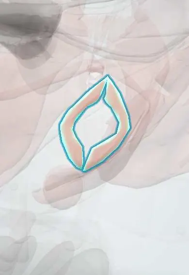
External anal sphincter !
The external anal sphincter is the ring of voluntary muscle around the anal canal; squeezing it keeps stool and gas in until it is time to empty the bowel.
Opens in the Durgical app, no sign-up needed.
What it is
A ring of voluntary muscle around the anal canal, in three layers.
What it does
Keeps stool and gas in until it is time to empty the bowel.
Key facts
- Origin
- Perineal body and skin around the anus (subcutaneous and superficial parts)
- Insertion
- Anococcygeal body; deep part encircles the upper anal canal, blending with puborectalis
- Action
- Voluntary closure of the anal canal
- Nerve
- Inferior rectal (anal) nerve from pudendal nerve (S2–S4)
- Blood supply
- Inferior rectal artery (internal pudendal)
Relations
- Medial
- Internal anal sphincter, separated by the intersphincteric space with the conjoint longitudinal muscle
- Lateral
- Fat of the ischioanal fossa, crossed by the inferior rectal nerve and vessels
- In front
- Perineal body; in women the lower vagina just ahead, in men the bulbospongiosus and the bulb of the penis
- Behind
- Anococcygeal body and the coccyx
- Above
- Puborectalis, with which its deep part fuses
How to find it
Its subcutaneous part surrounds the anal verge. On rectal examination the intersphincteric groove is felt between its lower edge and the internal sphincter; when the person squeezes, the ring tightens around the finger.
Examination
- Anal wink (anocutaneous reflex): stroking the perianal skin makes it contract; absence suggests a lesion of the S2–S4 segments or the pudendal nerve
- Rectal examination of squeeze: a weak or asymmetric squeeze suggests a sphincter defect or nerve injury
- Anorectal manometry: low squeeze pressure points to weakness of this muscle, low resting pressure to the internal sphincter
On imaging
Endoanal ultrasound shows it as a ring of mixed echogenicity outside the dark (hypoechoic) internal sphincter; a tear appears as a gap in the ring. Endoanal or pelvic MRI shows the sphincter layers and scarring.
Common problems
- Torn in a third- or fourth-degree perineal tear; if not repaired well it can cause faecal incontinence.
How it is diagnosed
Examination after birth; endoanal ultrasound if symptoms continue.
Treatment
- Repair by an experienced clinician right after birth; physiotherapy.
Clinical notes
- Anal fistulas are classified by their path through the sphincters (intersphincteric, transsphincteric, suprasphincteric, extrasphincteric); the more external sphincter a fistula crosses, the higher the risk of incontinence after surgery
- After a 3rd- or 4th-degree tear the torn ends are repaired end to end or overlapped; a persisting defect on ultrasound is linked to later incontinence
- Lateral internal sphincterotomy for chronic anal fissure divides the internal, not the external, sphincter
- Stretching of the pudendal nerve in a long second stage of labour can weaken it even without a visible tear
Anatomical variants
- Classic texts describe subcutaneous, superficial and deep parts; many modern studies find them hard to separate. In women the front of the ring is shorter than in men, which leaves less muscle in front to tear
Development
The anal canal below the anal membrane comes from the proctodeum, a pit of surface ectoderm, and the sphincter forms in the mesenchyme around it. In imperforate anus the sphincter is usually present at the normal site even though the bowel ends elsewhere; surgeons locate it with an electrical muscle stimulator and bring the rectum through it.
Related conditions
- Obstetric perineal tears and anal sphincter injury
- Haemorrhoids
- Anorectal abscess and fistula
- Rectal prolapse
- Fecal incontinence
- Anal fissure
- Anorectal malformations (imperforate anus)
Test yourself
Think of the answer, then tap the question to see it.
How are 3rd-degree perineal tears subclassified by the external anal sphincter?
3a: under half its thickness torn; 3b: over half; 3c: the internal anal sphincter is torn too. A 4th-degree tear also opens the anal mucosa.
Which sphincter gives most resting anal tone, and which the voluntary squeeze?
The internal anal sphincter (smooth, involuntary) gives resting tone; the external anal sphincter (striated, voluntary) gives the squeeze.
What does an absent anal wink (anocutaneous reflex) suggest?
A lesion of the S2–S4 segments or the pudendal nerve, which supply the external anal sphincter.
How do anal fistulas relate to the external anal sphincter and incontinence risk?
They are classified by path through the sphincters; the more external anal sphincter a fistula crosses, the higher the incontinence risk after surgery.
How does the external anal sphincter appear on endoanal ultrasound?
A ring of mixed echogenicity outside the dark internal anal sphincter; a tear appears as a gap in the ring.
Educational information, not medical advice. For any symptom, see a health professional.
Its names
- Official Latin (TA2)
- sphincter externus ani TA2 2426
- Official Spanish (DeCS)
- Músculo esfínter externo del ano DeCS D001003
- English (TA2)
- External anal sphincter
- Also called
- external anal sphincter muscle
Related
Sources: Terminologia Anatomica (TA2); DeCS; Anatomy, Abdomen and Pelvis: Anal Canal; Anatomy of the Human Body (20th ed.): The Digestive Apparatus (Lea & Febiger (Gray's Anatomy, 1918), 1918) · adapted, Public domain.
Spotted a mistake?We are always improving for the community. Tell us what is wrong and our team will review it.Report a mistake
Representative model, for educational purposes only.