Muscular system · Head and neck
Temporoparietalis !
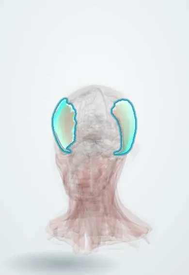
The temporoparietalis is a thin muscle over the side of the head above the ear; it helps tighten the scalp.
Official name: Temporoparietalis muscle
Opens in the Durgical app, no sign-up needed.
What it is
A thin muscle over the temple above the ear.
What it does
Tightens the scalp.
Key facts
- Origin
- Temporal fascia above the ear
- Insertion
- Epicranial aponeurosis
- Action
- Tightens the scalp
- Nerve
- Facial nerve (CN VII)
- Blood supply
- Superficial temporal and occipital arteries
Relations
- Superficial
- Subcutaneous tissue and skin of the temple, with the superficial temporal vessels and the auriculotemporal nerve
- Deep
- Loose areolar tissue, then the temporal fascia over the temporalis; the temporal branch of the facial nerve crosses here near the zygomatic arch
- Above
- Epicranial aponeurosis (galea)
- Below
- Continues as the SMAS of the face below the zygomatic arch
- Behind
- Anterior and superior auricular muscles above the auricle
How to find it
Not felt as a muscle: it is a thin, variable layer above and in front of the ear. In a dissection it is met right under the subcutaneous fat, with the pulsating superficial temporal artery on its surface.
On imaging
A thin sheet not usually seen as a separate muscle. On high-resolution ultrasound or MRI of the temple it lies within the temporoparietal fascia, with the superficial temporal artery and vein in the same layer. In giant cell arteritis, ultrasound of that artery looks for the halo sign, a dark ring of swollen wall. The fascia is also checked before use as a flap; the nerve branch is not seen.
Clinical notes
- The temporoparietal fascia flap, carried on the superficial temporal vessels, is a thin, well-vascularised flap used in ear reconstruction and to cover exposed structures in the head
- Superficial temporal artery biopsy for giant cell arteritis goes through the skin onto this layer
- In the coronal approach to the zygomatic arch the dissection drops deep to the superficial layer of the temporal fascia to keep the temporal branch safely in the flap
Anatomical variants
- Often thin, poorly developed or absent; texts treat it as part of the epicranius or of the auricular muscles
Development
Formed from the second (hyoid) arch muscle mass that spreads over the scalp and face, carrying the facial nerve; hence its supply by the temporal branch.
Test yourself
Think of the answer, then tap the question to see it.
In which fascial layer does the temporoparietalis sit?
The temporoparietal fascia, which joins the galea above to the SMAS below the zygomatic arch.
Why do surgeons dissect deep to the temporoparietal fascia over the zygomatic arch?
The temporal branch of the facial nerve runs on the deep side of this fascia; dissecting on the deep temporal fascia keeps the nerve safe in the flap.
Which vessels and nerve run within or on the temporoparietalis layer?
The superficial temporal artery and vein and the auriculotemporal nerve.
Educational information, not medical advice. For any symptom, see a health professional.
Its names
- Official Latin (TA2)
- musculus temporoparietalis TA2 2058
- Official Spanish (DeCS)
- Músculo temporoparietal
- English (TA2)
- Temporoparietalis muscle
Related
Sources: Terminologia Anatomica (TA2); Anatomy, Head and Neck: Facial Muscles; Anatomy, Head and Neck, Temporoparietal Fascia; Anatomy of the Human Body (20th ed.): Development of the Muscles (Lea & Febiger (Gray's Anatomy, 1918), 1918) · adapted, Public domain.
Spotted a mistake?We are always improving for the community. Tell us what is wrong and our team will review it.Report a mistake
Representative model, for educational purposes only.