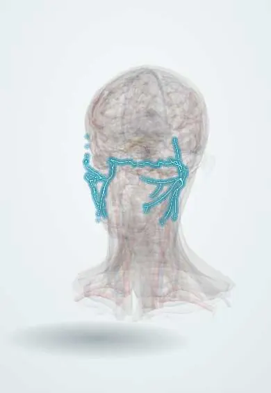
Nervous system · Head and neck
Facial nerve (VII) !
The facial nerve (cranial nerve VII) moves the muscles of facial expression, closes the eye and carries taste from the front of the tongue.
Official name: Facial nerve
Opens in the Durgical app, no sign-up needed.
What it is
The nerve that winds through the temporal bone, exits below the ear and fans across the face.
What it does
Moves the face, closes the eye, tastes the front of the tongue and drives tears and saliva.
Key facts
- Roots
- Motor nucleus (pons), superior salivatory, solitary nucleus; internal acoustic meatus -> facial canal -> stylomastoid foramen
- Moves
- Muscles of facial expression, stapedius, stylohyoid, posterior digastric; lacrimal, submandibular and sublingual glands (parasympathetic); branches: greater petrosal, nerve to stapedius, chorda tympani (facial canal); posterior auricular, digastric, stylohyoid; parotid plexus
- Feeling from
- Taste of the anterior two thirds of the tongue (chorda tympani); small area of the ear
Relations
- Course
- Leaves the pons at the cerebellopontine angle, enters the internal acoustic meatus, runs through the facial canal (bending at the geniculate ganglion, along the medial wall of the middle ear above the oval window, then down behind it) and exits at the stylomastoid foramen
- Below
- Below the stylomastoid foramen it gives the posterior auricular nerve and branches to the posterior belly of the digastric and the stylohyoid, then enters the parotid gland
- Superficial
- In the parotid gland it is the most superficial of the main structures, lying over the retromandibular vein and, deepest, the external carotid artery
- Lateral
- The middle ear cavity: the tympanic segment runs in its medial wall with the stapes and oval window just below and the lateral semicircular canal above and behind
How to find it
Its trunk is found in parotid surgery just deep and a little anterior and inferior to the tip of the tragal cartilage (tragal pointer), at the upper border of the posterior belly of the digastric and at the tympanomastoid suture, as it leaves the stylomastoid foramen.
Examination
- Motor exam: raise the eyebrows, close the eyes tightly, show the teeth, puff the cheeks; the House–Brackmann scale grades weakness from I (normal) to VI (complete palsy)
- Bell phenomenon: the eye rolls up and out when closure is attempted, visible when the lid fails to close
- Schirmer test (tear production), stapedial reflex and taste on the front of the tongue help place the lesion along the nerve
- Electroneurography and electromyography show how many fibres have degenerated and help predict recovery
On imaging
High-resolution temporal bone CT shows the facial canal (labyrinthine, tympanic, mastoid segments) and whether a fracture or cholesteatoma reaches it. MRI shows the cisternal, canalicular and parotid parts. Mild enhancement of the geniculate, tympanic and mastoid segments can be normal; in the meatus or labyrinthine segment it suggests Bell palsy or tumour. Typical Bell palsy needs no imaging.
Common problems
- Bell's palsy: sudden weakness of one whole side of the face, often after a virus.
- Stroke spares the forehead; Bell's palsy does not.
- Injury in parotid or ear surgery.
How it is diagnosed
Exam of forehead, eye closure and smile; tests are needed if the pattern is unusual.
Treatment
- Steroids started within 3 days; most people recover over weeks to months.
- Protecting the eye that cannot close (drops, ointment, taping at night).
Clinical notes
- Ramsay Hunt syndrome: herpes zoster of the geniculate ganglion with ear pain, vesicles in the concha or canal and facial palsy, sometimes with hearing loss or vertigo; recovery is poorer than in Bell palsy
- Temporal bone fractures, cholesteatoma and acute otitis media (through a dehiscent canal) can injure it inside the bone
- Facial weakness with a parotid lump suggests a malignant tumour; benign parotid tumours rarely cause palsy
- Faulty regeneration causes synkinesis (eye closure with smiling) and «crocodile tears» (tearing while eating)
- Eye protection is central in any palsy, because poor closure exposes the cornea
Anatomical variants
- The bony canal over the tympanic segment is often thin or open (dehiscent), leaving the nerve exposed in middle ear surgery and infection
Development
Nerve of the second pharyngeal arch, so it supplies that arch's muscles: facial expression, stapedius, stylohyoid and the posterior belly of the digastric.
Memory aid
What the facial nerve does: «face, ear, taste, tear» (facial muscles, stapedius, chorda tympani, lacrimal gland).
Related conditions
Test yourself
Think of the answer, then tap the question to see it.
Why is the forehead spared in a central (stroke) facial weakness but not in a peripheral facial nerve (VII) lesion?
The forehead motor neurons receive input from both hemispheres; a peripheral lesion paralyses the whole half of the face.
Which segment of the facial nerve is the narrowest, and why does it matter in Bell palsy?
The labyrinthine segment; swelling there chokes the nerve.
Which branches inside the temporal bone help localise a facial nerve lesion?
The greater petrosal nerve (tears), the nerve to stapedius (hyperacusis) and the chorda tympani (taste and saliva).
What is Ramsay Hunt syndrome?
Herpes zoster of the geniculate ganglion: ear pain, vesicles in the concha or canal and facial nerve palsy; recovery is poorer than in Bell palsy.
Through which foramen does the facial nerve leave the skull, and what is its landmark in parotid surgery?
The stylomastoid foramen; the trunk lies deep and slightly anteroinferior to the tragal pointer, at the upper border of the posterior digastric.
Educational information, not medical advice. For any symptom, see a health professional.
Its names
- Official Latin (TA2)
- nervus facialis TA2 6284
- Official Spanish (DeCS)
- Nervio Facial DeCS D005154
- English (TA2)
- Facial nerve
- Also called
- cranial nerve VII · nervus intermediofacialis
Related
- Internal acoustic meatus
- Stylomastoid foramen
- Parotid gland
- Frontalis
- Occipitalis
- Orbicularis oculi
- Orbicularis oris
- Buccinator
Sources: Terminologia Anatomica (TA2); DeCS; Facial Nerve Anatomy and Clinical Applications; Neuroanatomy, Geniculate Ganglion.
Spotted a mistake?We are always improving for the community. Tell us what is wrong and our team will review it.Report a mistake
Representative model, for educational purposes only.