Cardiovascular system · Head and neck
Superficial temporal artery !
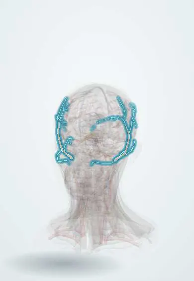
The superficial temporal artery feeds the scalp at the temple; its pulse is felt just in front of the ear, and it is sampled to diagnose giant cell arteritis.
Opens in the Durgical app, no sign-up needed.
What it is
The end of the external carotid, running up in front of the ear over the temple.
What it does
Feeds the scalp; its pulse is felt in front of the ear; it is biopsied for giant cell arteritis.
Key facts
- Origin
- Terminal branch of the external carotid in the parotid
- Branches
- Transverse facial, middle temporal, zygomatico-orbital, anterior auricular; frontal and parietal terminal branches
- Supplies
- Scalp and temple; pulse in front of the ear; biopsied in giant cell arteritis
Relations
- Course
- Begins in the parotid gland behind the neck of the mandible, crosses the posterior root of the zygomatic arch in front of the tragus and, a few centimetres higher, divides into frontal and parietal branches over the temple
- In front
- Temporal (frontal) branches of the facial nerve, crossing the zygomatic arch
- Behind
- Superficial temporal vein and auriculotemporal nerve
- Superficial
- Skin and the temporoparietal fascia, in which it runs
- Deep
- Root of the zygomatic arch, where the pulse is felt, then the temporal fascia and temporalis
How to find it
The pulse is felt just in front of the tragus, over the root of the zygomatic arch. In older people the frontal branch is often seen winding across the temple.
Examination
- Palpation in suspected giant cell arteritis: tenderness, thickening, nodules or a weak or absent pulse support the diagnosis
On imaging
Colour Doppler ultrasound in giant cell arteritis shows a dark ring of wall swelling around the lumen (halo sign) that stays visible when the probe compresses the artery (compression sign).
Clinical notes
- Visual loss in giant cell arteritis comes from inflamed branches of the ophthalmic artery (posterior ciliary arteries), not from the superficial temporal artery itself
- Superficial temporal to middle cerebral artery bypass (STA–MCA) is used in moyamoya disease and in some other blocked-artery situations
- The temporoparietal fascia flap, fed by this artery, is used to rebuild the ear and other parts of the face
- Pre-auricular incisions (parotid and temporomandibular joint surgery) pass next to it; it is either preserved or tied
Related conditions
Test yourself
Think of the answer, then tap the question to see it.
Where is the superficial temporal artery pulse felt?
Just in front of the tragus, over the root of the zygomatic arch.
What are the features of giant cell arteritis involving the superficial temporal artery?
Over age 50: new headache, scalp tenderness, jaw claudication and a thick, tender or pulseless artery; untreated, sudden permanent visual loss.
Why is a long segment taken in a superficial temporal artery biopsy, and which nerve is at risk?
The inflammation skips areas; the temporal (frontal) branch of the facial nerve is at risk.
Which bypass uses the superficial temporal artery, and for which disease?
The superficial temporal to middle cerebral artery (STA–MCA) bypass, used in moyamoya disease.
Educational information, not medical advice. For any symptom, see a health professional.
Its names
- Official Latin (TA2)
- arteria temporalis superficialis TA2 4413
- Official Spanish (DeCS)
- Arteria temporal superficial DeCS D013699
- English (TA2)
- Superficial temporal artery
Related
Sources: Terminologia Anatomica (TA2); DeCS; Anatomy, Head and Neck, Temporal Region; Anatomy, Head and Neck: Carotid Arteries.
Spotted a mistake?We are always improving for the community. Tell us what is wrong and our team will review it.Report a mistake
Representative model, for educational purposes only.