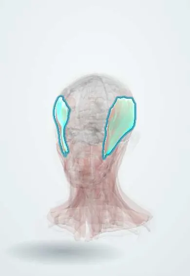
Muscular system · Head and neck
Temporalis !
The temporalis is a fan-shaped muscle on the side of the head under the temple; it closes the jaw and pulls it back.
Official name: Temporalis muscle
Opens in the Durgical app, no sign-up needed.
What it is
A fan-shaped muscle on the side of the head, under the temple.
What it does
Closes the jaw and pulls it back.
Key facts
- Origin
- Temporal fossa and temporal fascia
- Insertion
- Coronoid process and anterior ramus of the mandible
- Action
- Elevates the mandible; posterior fibres retract it
- Nerve
- Deep temporal nerves (V3)
- Blood supply
- Deep temporal arteries (maxillary artery), middle temporal artery
Relations
- Superficial
- The temporal fascia, then the temporoparietal fascia with the superficial temporal artery and auriculotemporal nerve, and the temporal branch of the facial nerve
- Deep
- Temporal fossa (frontal bone, parietal bones, greater wing of the sphenoid bone, squamous temporal bone) with the pterion; deep temporal vessels and nerves
- Below
- Passes deep to the zygomatic arch to the coronoid process; below, the lateral pterygoid and buccal fat pad in the infratemporal fossa
- Above
- Inferior temporal line, where its fibres begin
How to find it
With the fingertips on the temple above the zygomatic arch, the person clenches the teeth and the muscle hardens; its tendon can be felt on the coronoid process inside the mouth, along the front of the ramus.
Examination
- Clenching while both temples are palpated: loss of bulk or tightening on one side points to the mandibular nerve on that side
- Palpation of the muscle and its tendon for tender points in jaw pain and temporal headache
On imaging
CT and MRI show it filling the temporal fossa, deep to the temporal fascia, and narrowing to its tendon on the coronoid process. Contrast CT or MRI show a masticator space abscess spreading upward under the fascia, its suprazygomatic part. MRI shows wasting and fatty change after a mandibular nerve lesion. In trauma, CT shows the zygomatic arch, which it passes under, and coronoid fractures.
Clinical notes
- Hollowing of the temple after a pterional craniotomy comes from atrophy or retraction of the temporalis and loss of the fat pad
- The temporalis can be moved as a flap to fill the orbit or cover skull-base defects, and its tendon lengthened onto the lip for smile reanimation
- A fractured coronoid process is pulled up by the temporalis; coronoid enlargement can block mouth opening against the zygoma
- Jaw claudication and temporal tenderness in an older person raise concern for giant cell arteritis
Development
First pharyngeal arch muscle, supplied by the deep temporal nerves of V3.
Related conditions
- Tension-type headache
- Malnutrition and unintentional weight loss in older adults
- Myotonic dystrophy (types 1 and 2)
Test yourself
Think of the answer, then tap the question to see it.
What do the front and back fibres of the temporalis do?
Front fibres run vertically and close the jaw; back fibres run almost horizontally and pull the jaw back.
Which landmark and artery lie under the upper part of the temporalis?
The pterion, with the middle meningeal artery beneath.
Where does the temporalis insert, and what nerve supplies it?
On the coronoid process and anterior ramus of the mandible; deep temporal nerves (V3).
Why can the temple look hollow after a pterional craniotomy?
From atrophy or retraction of the temporalis and loss of the fat pad.
Educational information, not medical advice. For any symptom, see a health professional.
Its names
- Official Latin (TA2)
- musculus temporalis TA2 2108
- Official Spanish (DeCS)
- Músculo Temporal DeCS D013703
- English (TA2)
- Temporalis muscle
- Also called
- temporal muscle
Related
Sources: Terminologia Anatomica (TA2); DeCS; Anatomy, Head and Neck, Mastication Muscles; Anatomy, Head and Neck, Temporal Region.
Spotted a mistake?We are always improving for the community. Tell us what is wrong and our team will review it.Report a mistake
Representative model, for educational purposes only.