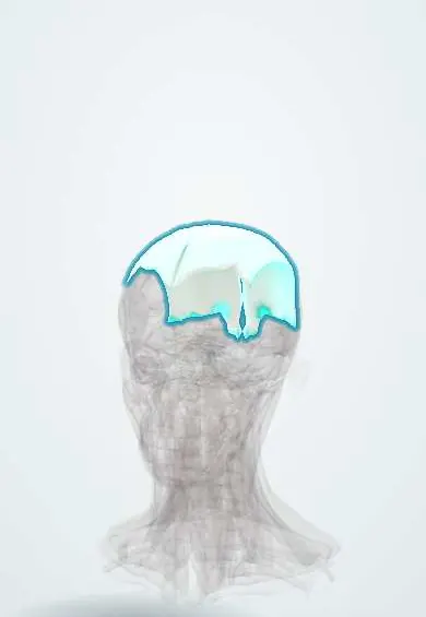
Muscular system · Head and neck
Galea !
The galea (epicranial aponeurosis) is a tough sheet over the top of the skull joining the forehead and back scalp muscles; scalp wounds through it gape open.
Official name: Epicranial aponeurosis
Opens in the Durgical app, no sign-up needed.
What it is
A tough sheet over the top of the skull joining the forehead and back scalp muscles.
What it does
Moves the scalp; scalp wounds gape when it is cut, and blood can spread beneath it.
Key facts
- Attaches
- Frontalis in front, occipitalis behind; temporal lines at the sides
- Function
- Moves the scalp; the 'A' layer of the scalp; wounds cut across it gape
Relations
- Superficial
- Dense connective tissue and skin, carrying the superficial temporal, supraorbital, supratrochlear, posterior auricular and occipital vessels and nerves
- Deep
- Loose areolar tissue, then the pericranium on the frontal bone, parietal bones and occipital bone
- In front
- Frontalis, which has no bony attachment and inserts into the skin of the brow
- Behind
- Occipitalis and, between the two bellies, attachment to the external occipital protuberance and highest nuchal lines
- Lateral
- Continues as the temporoparietal fascia (with the temporoparietalis) over the temporal fascia, reaching the superior temporal line and the zygomatic arch
How to find it
Not felt as a separate layer, but when the eyebrows are raised and lowered the whole scalp slides on the skull: the movement happens at the loose layer under the galea.
On imaging
On CT a subgaleal haematoma is a crescent over the skull that crosses suture lines; a cephalohaematoma (under the pericranium) stops at the sutures.
Clinical notes
- A laceration that cuts the galea gapes because frontalis and occipitalis pull the edges apart; repairing the galea also helps control bleeding
- Neonatal subgaleal haemorrhage, linked to vacuum delivery, can hold a large share of a baby's blood volume because the space runs from the orbits to the nuchal lines
- Blood or pus under the galea can track forward into the upper eyelids, because the frontalis inserts into skin rather than bone (bruised eyes after a scalp injury)
- Scalp flaps and the coronal approach to the skull are raised in the loose layer, the bloodless plane under the galea
Memory aid
SCALP: Skin, Connective tissue, Aponeurosis, Loose areolar tissue, Pericranium; the danger is in the L.
Related conditions
- Tension-type headache
- Dermatophytosis (tinea) and onychomycosis
- Seborrheic dermatitis
- Pediculosis (head, body and pubic lice)
- Alopecia areata
Test yourself
Think of the answer, then tap the question to see it.
What are the five layers of the scalp, and which is the epicranial aponeurosis?
Skin, dense connective tissue, aponeurosis (galea), loose areolar tissue and pericranium; the galea is the third, and the first three move as one.
Why is the loose areolar layer beneath the epicranial aponeurosis called the danger layer?
Emissary veins link it to the dural venous sinuses, so infection there can spread inside the skull.
Why does a scalp laceration that cuts the epicranial aponeurosis gape?
The frontalis and occipitalis pull the edges apart; repairing the galea also helps control bleeding.
How does a subgaleal haematoma differ from a cephalohaematoma on CT?
A subgaleal haematoma lies under the galea and crosses suture lines; a cephalohaematoma lies under the pericranium and stops at the sutures.
Educational information, not medical advice. For any symptom, see a health professional.
Its names
- Official Latin (TA2)
- galea aponeurotica TA2 2059
- Official Spanish (DeCS)
- Aponeurosis epicraneal
- English (TA2)
- Epicranial aponeurosis
Sources: Terminologia Anatomica (TA2); Anatomy, Head and Neck, Scalp; Anatomy, Head and Neck; Frontalis Muscle; Tratado de anatomía humana (9.ª ed.), tomo II, Libro IV Angiología, sección 5: Linfáticos.
Spotted a mistake?We are always improving for the community. Tell us what is wrong and our team will review it.Report a mistake
Representative model, for educational purposes only.