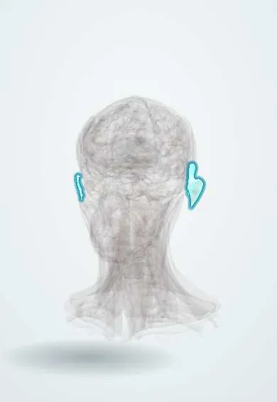
Auricle !
The auricle is the visible outer ear, skin over a shaped plate of elastic cartilage; it collects sound and funnels it into the ear canal.
Opens in the Durgical app, no sign-up needed.
What it is
The visible ear: skin over a shaped plate of elastic cartilage.
What it does
Collects sound and funnels it into the ear canal.
Key facts
- Where
- Side of the head; elastic cartilage (except the lobule)
- Blood supply
- Posterior auricular and superficial temporal arteries
- Nerve
- Great auricular (C2–C3), auriculotemporal (V3), lesser occipital; VII/X small areas
- Function
- Collects sound
Relations
- In front
- Tragus, overlapping the opening of the external acoustic meatus; just in front, the superficial temporal artery, the auriculotemporal nerve and the parotid gland
- Behind
- Retroauricular groove over the mastoid process of the temporal bone, with the posterior auricular artery and branches of the great auricular nerve
- Medial
- The concha leads into the cartilaginous lateral part of the ear canal, whose cartilage is continuous with that of the auricle
- Superficial
- Thin skin bound tightly to the perichondrium on the lateral surface, more loosely on the back; little room for swelling, so lesions on the front are painful
How to find it
The whole outline is visible: the rim (helix) curls forward into the crus of the helix above the concha; the antihelix runs parallel inside it and splits above into two crura around the triangular fossa. The tragus sits in front of the canal opening and the lobule hangs below. Behind the ear the mastoid process is felt in the retroauricular groove.
Examination
- Inspection behind the ear: a tender retroauricular swelling that pushes the auricle forward and outward suggests acute mastoiditis complicating otitis media
- Pain on pressing the tragus or pulling the auricle points to otitis externa; it is usually absent in otitis media
On imaging
Usually judged by eye. Ultrasound can confirm fluid under the perichondrium in an auricular haematoma or perichondritis. When infection or a skin cancer spreads from the auricle toward the canal and skull base, CT shows bone erosion and MRI the soft tissue spread. In microtia, high-resolution temporal bone CT assesses the canal, middle ear and facial nerve before reconstruction.
Clinical notes
- Perichondritis, typically after piercing through cartilage or burns (Pseudomonas is the classic organism): red, painful, swollen auricle that spares the lobule, which has no cartilage
- The sun-exposed helix is a frequent site of actinic keratosis, basal and squamous cell carcinoma, and of frostbite
- Conchal cartilage and tragal cartilage with perichondrium are standard autologous grafts (rhinoplasty, eardrum repair)
- Vesicles in the concha and canal with facial weakness suggest herpes zoster of the geniculate ganglion (Ramsay Hunt syndrome)
Anatomical variants
- Preauricular pit or sinus just in front of the root of the helix, from incomplete fusion of the hillocks; it can become infected and form abscesses
- Preauricular skin tags (accessory auricles) along the line from the tragus to the angle of the mouth
- Darwin tubercle: a small knob on the upper back part of the helix
- Prominent ears from an underdeveloped antihelical fold or a deep concha
Development
Forms from six auricular hillocks around the first pharyngeal cleft, three from the first arch and three from the second; the ear starts low on the neck and rises, so low-set or malformed ears can flag genetic syndromes and middle-ear anomalies.
Related conditions
- Otitis externa
- Cutaneous squamous cell carcinoma
- Discoid lupus erythematosus
- Keloid and hypertrophic scar
- Relapsing polychondritis
Test yourself
Think of the answer, then tap the question to see it.
Why can an untreated auricular haematoma lead to «cauliflower ear»?
The cartilage depends on the perichondrium for nutrition; blood lifting it off starves the cartilage, and the clot organises into fibrous, deformed tissue.
Why does perichondritis of the auricle spare the lobule?
The lobule has no cartilage; perichondritis follows piercing through cartilage or burns, classically with Pseudomonas.
What does a tender retroauricular swelling pushing the auricle forward and outward suggest?
Acute mastoiditis complicating otitis media.
What do vesicles in the concha and ear canal with facial weakness suggest?
Herpes zoster of the geniculate ganglion (Ramsay Hunt syndrome).
Educational information, not medical advice. For any symptom, see a health professional.
Its names
- Official Latin (TA2)
- auricula TA2 104
- Official Spanish (DeCS)
- Pabellón Auricular DeCS D054644
- English (TA2)
- Auricle
- Also called
- pinna · outer ear
Related
Sources: Terminologia Anatomica (TA2); DeCS; Ear, Hearing, and Facial Nerve Evaluation; Auricular Hematoma.
Spotted a mistake?We are always improving for the community. Tell us what is wrong and our team will review it.Report a mistake
Representative model, for educational purposes only.