Nervous system · Head and neck
Hypoglossal nerve (XII) !
The hypoglossal nerve (cranial nerve XII) moves the tongue; if one side is damaged, the tongue points toward that side when stuck out.
Official name: Hypoglossal nerve
Opens in the Durgical app, no sign-up needed.
What it is
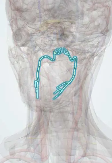
The nerve from the medulla that loops forward under the jaw to the tongue.
What it does
Moves the tongue; when one side is damaged the tongue points to that side.
Key facts
- Roots
- Hypoglossal nucleus (medulla); hypoglossal canal
- Moves
- All tongue muscles except palatoglossus (C1 fibres ride along to geniohyoid, thyrohyoid)
- Feeling from
- None
Relations
- Course
- Leaves the medulla oblongata as rootlets between the pyramid and the olive, exits through the hypoglossal canal, descends between the internal carotid artery and the internal jugular vein, curves forward over both carotid arteries below the posterior belly of the digastric, then runs on the hyoglossus into the tongue
- Superficial
- The posterior belly of the digastric where it crosses the carotids, then the mylohyoid in the floor of the mouth
- Deep
- The internal and external carotid arteries as it crosses them, then the hyoglossus, which separates it from the lingual artery
- Above
- On the hyoglossus, the lingual nerve and the submandibular duct
How to find it
In the neck it is found just above the carotid bifurcation, crossing the carotids under the posterior belly of the digastric, often with a sternocleidomastoid branch of the occipital artery hooking over it; its descending branch (C1 fibres) leads down to the ansa cervicalis.
Examination
- Tongue at rest in the mouth: wasting and fasciculations point to a lower motor neuron lesion
- Tongue protrusion: deviation to one side means weakness of that side (lower motor neuron) or of the opposite hemisphere (upper motor neuron)
- Pushing the tongue into each cheek against the examiner's finger compares strength side to side
On imaging
T2-weighted and contrast MRI follow it from the medulla, in front of the olive, through the hypoglossal canal; bone-window CT shows the canal itself. A palsy leaves half of the tongue atrophied and fatty; early on that half may look swollen and bright on MRI and mimic tumour. Imaging then traces the nerve back through the carotid space and canal for a cause, such as carotid dissection.
Clinical notes
- Wasting and fasciculations on both sides suggest motor neuron disease with bulbar onset
- Medial medullary syndrome: tongue weakness on the same side with weakness of the opposite side of the body
- Carotid artery dissection and skull base lesions near the hypoglossal canal can cause an isolated palsy
- The nerve can be transferred to the facial nerve (VII) to restore facial movement after facial nerve loss
- Upper side of two surgical triangles: Farabeuf (with the internal jugular vein and thyrolinguofacial trunk), over the carotid bifurcation, and Pirogoff (with the digastric tendon and mylohyoid), over the lingual artery
Development
The tongue muscles come from occipital somites, not from a pharyngeal arch, which is why they have their own nerve.
Memory aid
"Lick your wounds": in a nerve lesion the tongue points toward the damaged side.
Related conditions
Test yourself
Think of the answer, then tap the question to see it.
Which way does the protruded tongue deviate in a lower motor neuron hypoglossal nerve (XII) lesion, and why?
Toward the side of the lesion, pushed by the healthy genioglossus, with wasting and fasciculations on that side.
How does the tongue deviate in an upper motor neuron lesion such as a stroke?
Away from the side of the brain lesion, without wasting.
Where is the hypoglossal nerve at risk in carotid endarterectomy?
Just above the carotid bifurcation, where it crosses the internal and external carotid arteries under the posterior belly of the digastric.
What is medial medullary syndrome?
Tongue weakness on the same side (hypoglossal nerve) with weakness of the opposite side of the body.
Educational information, not medical advice. For any symptom, see a health professional.
Its names
- Official Latin (TA2)
- nervus hypoglossus TA2 6357
- Official Spanish (DeCS)
- Nervio Hipogloso DeCS D007002
- English (TA2)
- Hypoglossal nerve
- Also called
- cranial nerve XII
Related
Sources: Terminologia Anatomica (TA2); DeCS; Neuroanatomy, Cranial Nerve 12 (Hypoglossal); Anatomía humana (4.ª ed.), tomo 1, cap. 36: Nervios craneales.
Spotted a mistake?We are always improving for the community. Tell us what is wrong and our team will review it.Report a mistake
Representative model, for educational purposes only.