Conditions · Metabolism: carbohydrates and glycogen
Classic galactosaemia !
In classic galactosaemia the baby cannot process milk galactose: within days vomiting, jaundice and liver damage appear, and it can be fatal.
Updated 2026-11-01
Structures involved
Tap one to see what it is and open it in 3D.
In biochemistry
- Galactose metabolism: Step 3 · Galactose-1-phosphate uridylyltransferase (GALT)
- Galactose-1-phosphate uridylyltransferase (GALT)
- UDP-glucose
- Galactose
- Carbohydrate chemistry
- Newborn screening
In physiology
What it is
Galactosaemia type I: near-total lack of galactose-1-phosphate uridylyltransferase (GALT). It is a newborn emergency sought on newborn screening.
Who gets it
About 1 in 30,000 to 60,000 newborns. In Colombia expanded newborn screening includes it in some programmes, but not nationwide.
Causes
- Autosomal recessive inheritance: GALT gene variants (Q188R commonest in people of European descent)
Risk factors
- Carrier parents or affected siblings
- Feeding with breast milk or lactose-containing formula (trigger)
Pathophysiology
Without GALT, galactose 1-phosphate builds up in liver, kidney, brain and lens, where it is toxic. Free galactose is reduced to galactitol (aldose reductase), which draws water into the lens and clouds it, and oxidised to galactonate. Liver damage causes jaundice and coagulopathy; tubular damage, aminoaciduria. High galactose impairs neutrophil function: risk of E. coli sepsis.
Classification
- Classic (type I): almost no GALT activity
- Clinical variant: some residual activity, commoner in people of African descent
- Duarte variant: mild, usually symptom-free
Symptoms and signs
- Poor feeding and vomiting in the first days of life
- Jaundice and hepatomegaly
- Lethargy and hypotonia
- Cataract
- Bleeding from coagulopathy
Diagnosis
- Clinical
- Newborn who worsens on milk feeds, with jaundice, hepatomegaly and sometimes sepsis or cataract.
- Tests
- Newborn screening (GALT activity or total galactose in dried blood). Confirmation: red cell GALT activity, red cell galactose 1-phosphate and GALT gene testing. Reducing sugars in urine; abnormal transaminases, bilirubin and clotting. A recent transfusion can mislead GALT activity.
- Imaging
- Slit-lamp examination for cataract; abdominal ultrasound for the liver.
Treatment
- Acute
- Stop breast milk and lactose-containing formula immediately on suspicion, without waiting for confirmation
- Lifestyle
- Lactose-free formula (for example soy-based) and a lifelong lactose- and galactose-free diet
- Acute
- Treatment of sepsis, coagulopathy and liver failure at presentation
- Follow-up
- Follow-up of development, speech, eyes, bone density and ovarian function
Complications
- Neonatal E. coli sepsis
- Liver failure
- Speech and learning delay
- Primary ovarian insufficiency
- Cataract
Prognosis
Early removal of galactose saves life and reverses liver damage and cataract; even so, speech and learning problems and ovarian failure can remain.
Prevention
- Newborn screening and genetic counselling for families
Test yourself
Think of the answer, then tap the question to see it.
Which enzyme is missing in classic galactosaemia?
Galactose-1-phosphate uridylyltransferase (GALT).
Which infection is typical in the newborn with galactosaemia?
Escherichia coli sepsis.
What is done when galactosaemia is suspected in a newborn?
Stop lactose immediately (breast milk and lactose formula) without waiting for confirmation.
Which molecule causes cataract in galactosaemia?
Galactitol, formed by aldose reductase from galactose.
Related conditions
Educational information, not medical advice. For any symptom, see a health professional.
Sources: Galactosemia: MedlinePlus Genetics (MedlinePlus, US National Library of Medicine, 2026); Galactosemia: MedlinePlus Medical Encyclopedia (MedlinePlus, US National Library of Medicine, 2026); GALT gene: MedlinePlus Genetics (MedlinePlus, US National Library of Medicine, 2026); Textbook of Biochemistry for Medical Students, 6th ed. (Jaypee Brothers Medical Publishers (Vasudevan DM, Sreekumari S, Vaidyanathan K), 2011).
Spotted a mistake?We are always improving for the community. Tell us what is wrong and our team will review it.Report a mistake
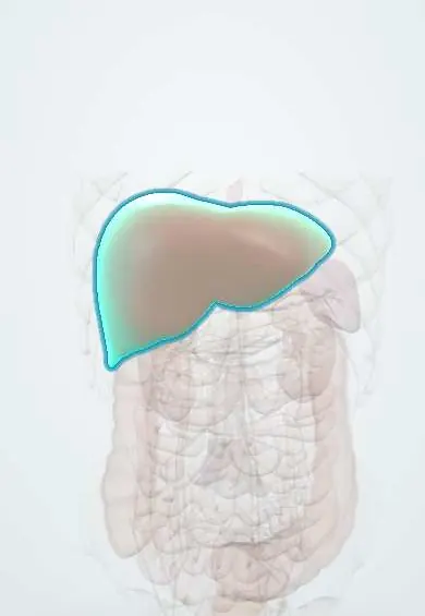
Representative model, for educational purposes only.