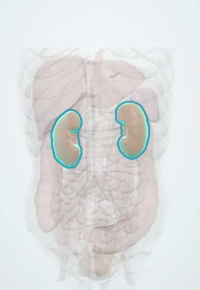
Kidneys !
The kidneys are two bean-shaped organs at the back of the abdomen, on either side of the spine; they filter the blood and make urine.
Official name: Kidney
Opens in the Durgical app, no sign-up needed.
What it is
Two bean-shaped organs against the back wall of the abdomen, beside the spine at about T12-L3; the right sits a little lower because of the liver.
What it does
Filter about 180 litres of blood plasma a day to make 1-2 litres of urine, balancing water, salts and acid, and make hormones that control blood pressure and red-cell production.
Key facts
- Where
- Retroperitoneal, T12–L3, right a little lower; hilum at L1 (transpyloric plane); inside renal (Gerota) fascia with perirenal fat
- Blood supply
- Renal arteries from aorta (L1–L2); renal veins into IVC
- Nerve
- Renal plexus: sympathetic T10–L1 (lesser, least and lumbar splanchnics), vagal fibres; pain from loin to groin
- Function
- Filter plasma and make urine; water, electrolyte and acid–base balance; renin, erythropoietin, active vitamin D
Relations
- In front
- Right: right adrenal gland, liver across the hepatorenal pouch, second part of the duodenum, hepatic flexure of the colon, small intestine. Left: left adrenal gland, stomach, spleen, tail of the pancreas with the splenic vessels, splenic flexure and descending colon, jejunum
- Behind
- Diaphragm above, with the pleural costodiaphragmatic recess behind it; psoas major medially, quadratus lumborum in the middle, transversus abdominis laterally
- Above
- Adrenal gland on the upper pole, in its own compartment of the renal (Gerota) fascia
- Medial
- Hilum with the renal vessels and pelvis; then the inferior vena cava on the right and the abdominal aorta on the left
How to find it
The renal angle lies between the 12th rib and the lateral edge of the erector spinae. The kidneys are felt bimanually, one hand in the loin pushing forward and the other in the flank; the lower pole of the right kidney may be felt in a thin person at the end of a deep breath.
Examination
- Renal angle (costovertebral) tenderness on gentle percussion: points to pyelonephritis, an obstructing stone or a perinephric abscess
- Ballottement: a mass bounced forward between the two hands is renal; unlike the spleen it is resonant in front (colon), has no notch and the hand can get above it
On imaging
Ultrasound is the first test for size, cortical thickness, hydronephrosis and cysts. Non-contrast CT shows stones; CT urography (non-contrast, nephrographic and excretory phases) studies hematuria and urothelial lesions. On a trauma FAST scan, free fluid is looked for first in the hepatorenal pouch (Morison's pouch), between the liver and the right kidney.
Common problems
- Kidney stones.
- Chronic kidney disease, mostly from diabetes and high blood pressure.
- Infections (pyelonephritis).
- Cysts and tumours.
How it is diagnosed
Blood (creatinine) and urine tests; ultrasound; CT.
Treatment
- Control blood pressure and diabetes; treat infections; dialysis or a kidney transplant for kidney failure.
Clinical notes
- The pleura dips below the 12th rib medially, so posterior access to the upper pole (percutaneous nephrolithotomy, biopsy, surgery) can cause a pneumothorax
- Renal cell carcinoma can grow into the renal vein and up the inferior vena cava; a left varicocele that appears in an older man raises the question of a left renal tumour
- Pyelonephritis usually spreads up from the bladder; vesicoureteric reflux is an important risk in children
- Living kidney donation: the left kidney is usually chosen because its renal vein is longer
Anatomical variants
- Horseshoe kidney: the lower poles fuse across the midline and the kidney is held low because its isthmus catches under the inferior mesenteric artery; the commonest fusion anomaly
- Duplex collecting system (two pelves and ureters on one side) and ectopic, often pelvic, kidney
Development
The metanephros forms from the ureteric bud and the metanephric blastema in the pelvis and climbs to the loin, taking blood from successively higher arteries: the origin of accessory renal arteries and of low or fused kidneys.
Related conditions
- Kidney stones (urolithiasis)
- Acute pyelonephritis
- Chronic kidney disease
- Acute kidney injury
- Autosomal dominant polycystic kidney disease
- Arsenic poisoning
- Lactic acidosis
- Diabetic ketoacidosis
- Tyrosinaemia type I (hepatorenal)
- Cystinuria
- Hartnup disease
- Methylmalonic acidaemia
- Von Gierke disease (glycogen storage disease type I)
- Hereditary fructose intolerance
- Essential fructosuria
- Fabry disease
- High anion gap metabolic acidosis
- Normal anion gap (hyperchloraemic) metabolic acidosis
- Metabolic alkalosis
- Hyponatraemia
- Hypernatraemia
- Hypokalaemia
- Hyperkalaemia
- Hypercalcaemia
- Primary hyperoxaluria
- Lead poisoning
- Fanconi anaemia
- Lesch-Nyhan syndrome
- Hereditary orotic aciduria
- Tumour lysis syndrome
- Hereditary xanthinuria
- Amyloidosis
- Ethylene glycol poisoning
- Mercury poisoning
- Cadmium poisoning
- Type 2 diabetes mellitus
- Type 1 diabetes mellitus
- Arterial hypertension
- Sepsis and septic shock
- Systemic lupus erythematosus
- Hypovolaemic shock
- Leptospirosis
- Snakebite envenomation (Bothrops, Lachesis, Crotalus)
- Salicylate (aspirin) toxicity
- Heat stroke and heat exhaustion
- Paraquat poisoning
- Spider bites (loxoscelism and latrodectism)
- Anticoagulant poisoning (warfarin and superwarfarins)
- Mushroom poisoning (Amanita)
- Diabetes insipidus (central and nephrogenic)
- Hyperosmolar hyperglycaemic state
- Polypharmacy and potentially inappropriate medications in older adults
- Hepatorenal syndrome
- Disseminated intravascular coagulation
- Transfusion reactions
- Thrombotic thrombocytopenic purpura
- Anaemia of chronic disease (anaemia of inflammation)
- Hypercalcaemia of malignancy
- Paroxysmal nocturnal haemoglobinuria
- Yellow fever
- Nephrotic syndrome
- Post-streptococcal glomerulonephritis
- Diabetic kidney disease
- Dehydration (fluid volume deficit)
- Lupus nephritis
- Minimal change disease
- Focal segmental glomerulosclerosis
- Membranous nephropathy
- Renal tubular acidosis
- Acute interstitial nephritis
- Alport syndrome
- CKD-mineral and bone disorder (renal osteodystrophy)
- Renal cell carcinoma
- Fluid volume overload (hypervolaemia)
- IgA nephropathy
- Rapidly progressive glomerulonephritis (anti-GBM, ANCA)
- Rhabdomyolysis
- Renovascular hypertension (renal artery stenosis)
- Hypomagnesaemia
- Vesicoureteral reflux
- Kidney transplant rejection
- Membranoproliferative glomerulonephritis
- Hypertensive nephrosclerosis
- Hypophosphataemia
- Fanconi syndrome
- Bartter, Gitelman and Liddle syndromes
- Congenital kidney anomalies (horseshoe kidney, renal agenesis)
- Renal papillary necrosis
- Medullary sponge kidney and medullary cystic disease
- Asymptomatic bacteriuria
- Calciphylaxis (calcific uremic arteriolopathy)
- Atheroembolic renal disease (cholesterol crystal embolism)
- Dehydration in children (acute diarrhoeal disease)
- Wilms tumour (nephroblastoma)
- Hemolytic uremic syndrome (HUS)
- Beckwith–Wiedemann syndrome
- VACTERL association
- Complement deficiencies
- Autosomal recessive polycystic kidney disease
- Lithium toxicity
- Systemic sclerosis (scleroderma, including CREST)
- Antiphospholipid syndrome
- Granulomatosis with polyangiitis
- IgA vasculitis (Henoch–Schönlein purpura)
- Microscopic polyangiitis
- Cryoglobulinemic vasculitis
- Familial Mediterranean fever and other hereditary autoinflammatory syndromes
- Blunt abdominal trauma
- Renal trauma
- Abdominal compartment syndrome
Test yourself
Think of the answer, then tap the question to see it.
What is the order of structures at the renal hilum, from front to back?
Renal vein, renal artery, renal pelvis (VAP).
At what vertebral levels do the kidneys lie, and why is the right one lower?
Retroperitoneal at about T12–L3, with the hilum at L1 (transpyloric plane); the right kidney sits a little lower because of the liver.
Why can posterior access to the upper pole of the kidney cause a pneumothorax?
The pleura dips below the 12th rib medially, so percutaneous nephrolithotomy, biopsy or surgery at the upper pole can enter the pleural space.
What holds a horseshoe kidney low in the abdomen?
Its isthmus, formed by the fused lower poles, catches under the inferior mesenteric artery; it is the commonest renal fusion anomaly.
Why is the left kidney usually chosen for living kidney donation?
The left renal vein is longer, which makes the transplant join easier.
Educational information, not medical advice. For any symptom, see a health professional.
Its names
- Official Latin (TA2)
- ren; cortex renis; medulla renis TA2 3358
- Official Spanish (DeCS)
- Riñón DeCS D007668
- English (TA2)
- Kidney
Related
Sources: Terminologia Anatomica (TA2); DeCS; Anatomy, Abdomen and Pelvis: Kidneys.
Spotted a mistake?We are always improving for the community. Tell us what is wrong and our team will review it.Report a mistake
Representative model, for educational purposes only.