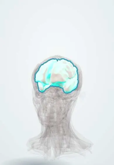
Nervous system · Head and neck
White matter !
The white matter is the brain's wiring under the cortex: bundles of nerve fibres that link brain areas with each other and with the spinal cord.
Official name: White matter of telencephalon
Opens in the Durgical app, no sign-up needed.
What it is
The wiring under the cortex: bundles of nerve fibres, including the internal capsule.
What it does
Connects cortex areas with each other and with the brainstem and spinal cord.
Key facts
- Where
- Beneath the cortex; internal capsule between thalamus/caudate and lentiform nucleus
- Blood supply
- Cortical (medullary) branches of the ACA, MCA and PCA; internal capsule: lenticulostriate arteries (MCA, most), recurrent artery of Heubner (anterior limb), anterior choroidal artery (lower posterior limb and retrolenticular part)
- Function
- Connections; internal capsule carries corticospinal fibres (small strokes = big deficits)
Relations
- Superficial
- Cerebral cortex, with short U-fibres linking neighbouring gyri just beneath it
- Deep
- Lateral ventricles; the caudate nucleus, thalamus and lentiform nucleus lie embedded in it
- Contents
- Corona radiata, centrum semiovale, internal, external and extreme capsules; long association bundles such as the arcuate (superior longitudinal), uncinate, cingulum and inferior longitudinal fasciculi
- Course
- Corticospinal fibres converge from the motor cortex through the corona radiata into the posterior limb of the internal capsule, then into the cerebral peduncle
How to find it
On axial MRI the centrum semiovale is the white matter above the ventricles, the corona radiata lies at the level of the ventricle bodies, and the internal capsule forms a V at the basal ganglia level, with its genu pointing toward the midline.
On imaging
In adults white matter is bright on T1 and dark on T2; in newborns the pattern is reversed until myelination advances. FLAIR shows plaques and small-vessel disease, susceptibility-weighted images show the microbleeds of axonal injury, and diffusion tensor imaging traces the tracts.
Clinical notes
- Lenticulostriate branches of the middle cerebral artery supply the internal capsule; long-standing high blood pressure damages them, causing lacunes and haemorrhage
- Classic lacunar syndromes: pure motor (internal capsule), pure sensory (thalamus), ataxic hemiparesis and dysarthria–clumsy hand
- Widespread small-vessel white matter disease is linked to slowed thinking, gait problems and urinary urgency in older adults
- Anterior choroidal artery infarct: posterior limb of the internal capsule, optic tract and lateral geniculate body, giving opposite-side weakness, numbness and homonymous hemianopia
- Deep perforating arteries and the long medullary branches from the cortex do not join, so the white matter between them (centrum semiovale, beside the ventricles) is a border zone where small-vessel and low-flow infarcts gather
Development
Myelination runs from back to front and from deep to superficial: the posterior limb of the internal capsule is myelinated around birth, while frontal association tracts keep maturing into early adulthood.
Related conditions
- Phenylketonuria (PKU)
- Maple syrup urine disease
- Ornithine transcarbamylase (OTC) deficiency
- Hyperammonaemia
- Krabbe disease (globoid cell leukodystrophy)
- Metachromatic leukodystrophy
- Zellweger syndrome (Zellweger spectrum)
- X-linked adrenoleukodystrophy
- Pyruvate carboxylase deficiency
- Glycine encephalopathy (nonketotic hyperglycinaemia)
- Guanidinoacetate methyltransferase (GAMT) deficiency
- Sanfilippo syndrome (mucopolysaccharidosis type III)
- Lead poisoning
- Carbon monoxide poisoning
- Menkes disease
- Multiple sclerosis
- Glioblastoma
- Zika virus infection
- High-altitude illness (AMS, HACE, HAPE)
- Dementia (major neurocognitive disorder)
- Primary central nervous system lymphoma
- Nocardiosis
- Vascular dementia
- Brain metastases
- Eclampsia
- Perinatal asphyxia and hypoxic-ischaemic encephalopathy
- Congenital (TORCH) infections: syphilis, toxoplasmosis, CMV and Zika
- Cerebral palsy
- Reye syndrome
- Infantile spasms (West syndrome)
Test yourself
Think of the answer, then tap the question to see it.
What are the three types of fibres in the cerebral white matter?
Projection (cortex to and from the thalamus, brainstem and cord), association (within one hemisphere) and commissural (between hemispheres, such as the corpus callosum).
Why does a small lacune in the posterior limb of the internal capsule cause a large deficit?
Corticospinal fibres for the arm and leg are packed there; a small lacune causes pure motor hemiparesis.
Which arteries supply the internal capsule, and what does long-standing high blood pressure do to them?
Lenticulostriate branches of the middle cerebral artery; hypertension damages them, causing lacunes and haemorrhage.
Where do multiple sclerosis plaques favour in the white matter?
Periventricular, juxtacortical and callosal white matter, often ovoid and perpendicular to the ventricles (Dawson fingers).
What is diffuse axonal injury?
Shearing from acceleration–deceleration at the grey–white junction, corpus callosum and brainstem; coma is out of proportion to the CT.
Educational information, not medical advice. For any symptom, see a health professional.
Its names
- Official Latin (TA2)
- substantia alba telencephali TA2 5573
- Official Spanish (DeCS)
- Sustancia blanca del telencéfalo DeCS D066127
- English (TA2)
- White matter of telencephalon
Sources: Terminologia Anatomica (TA2); DeCS; Neuroanatomy, Internal Capsule; Neuroanatomy, Gray Matter; Anatomía humana (4.ª ed.), tomo 1, cap. 32: Configuración interna y sistematización del prosencéfalo; Anatomía humana (4.ª ed.), tomo 1, cap. 26: Vascularización del encéfalo; Tratado de anatomía humana (9.ª ed.), tomo II, Sistema nervioso central: cerebro, núcleos grises centrales, vías de conducción y circulación cerebral.
Spotted a mistake?We are always improving for the community. Tell us what is wrong and our team will review it.Report a mistake
Representative model, for educational purposes only.