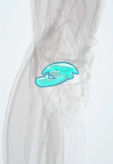
Tibial cartilage !
The tibial cartilage is the smooth layer covering the top of the shin bone inside the knee; it lets the joint glide and spreads load.
Official name: Articular cartilage of tibial plateau
Opens in the Durgical app, no sign-up needed.
What it is
Articular cartilage covering the top of the tibia, partly under the menisci.
What it does
Lets the joint glide with very little friction and spreads load across the bone.
Key facts
- Where
- Medial and lateral tibial plateaus, partly covered by the menisci
- Function
- Hyaline articular cartilage; low-friction gliding; spreads load; avascular, nourished by synovial fluid
Relations
- Above
- Femoral condyle cartilage; the menisci over the outer part of each plateau
- In front
- Anterior cruciate ligament footprint and the anterior meniscal horns in front of the intercondylar eminence; infrapatellar fat pad
- Behind
- Posterior cruciate ligament footprint and the posterior meniscal horns behind the plateaus
- Deep
- Subchondral bone of the plateaus
How to find it
The tibiofemoral joint line can always be felt. With the knee slightly bent it lies just behind the lower tip of the patella; on the medial side a small triangular hollow, framed by the edge of the patella, the medial femoral condyle and the top of the medial tibial condyle, marks the joint. At arthroscopy the plateau is probed beneath the femoral condyle with the knee in varus or valgus.
On imaging
Standing and Rosenberg views for joint-space narrowing; long-leg standing views show varus or valgus load. MRI shows cartilage loss and bone marrow lesions. In plateau fractures, CT measures the step and depression of the joint surface.
Common problems
- Cartilage injuries (focal defects) from sports or a twisting injury.
- Osteoarthritis: gradual wear of the cartilage, causing pain and stiffness.
How it is diagnosed
Symptoms and exam; X-rays show joint-space narrowing in arthritis; MRI shows cartilage defects.
Treatment
- Physical therapy, weight management, activity changes; medicines or injections for pain.
- Surgery for focal defects: microfracture, cell-based repair, or osteochondral grafts (including osteochondral allograft from a donor).
- Knee replacement for advanced arthritis.
Clinical notes
- A residual step in the joint surface after a plateau fracture leads to post-traumatic arthritis
- High tibial osteotomy shifts load from a worn medial compartment to the healthier lateral one in younger, active people
- Unicompartmental knee replacement resurfaces only the worn compartment when the rest of the joint is preserved
Development
Retained hyaline cartilage of the embryonic model of the upper tibia; the joint cavity appears when the mesenchyme between femur and tibia loosens. The proximal tibial epiphysis starts to ossify around birth.
Related conditions
Test yourself
Think of the answer, then tap the question to see it.
Which compartment of the knee is the commonest site of osteoarthritis, and with what alignment?
The medial compartment, linked to varus alignment.
What does chondrocalcinosis look like on a knee X-ray?
Fine lines of calcium pyrophosphate within the tibial cartilage and menisci.
What is the aim of a high tibial osteotomy?
To shift load from a worn medial compartment to the healthier lateral one, in younger, active people.
Educational information, not medical advice. For any symptom, see a health professional.
Its names
- Official Latin (TA2)
- cartilago articularis TA2 382
- Official Spanish (DeCS)
- Cartílago articular de la tibia (platillos) DeCS D002358
- English (TA2)
- Articular cartilage of tibial plateau
Related
Sources: Terminologia Anatomica (TA2); DeCS; Anatomy, Bony Pelvis and Lower Limb, Knee; Anatomy of the Human Body (20th ed.): Surface Anatomy of the Lower Extremity (Lea & Febiger (Gray's Anatomy, 1918), 1918) · adapted, Public domain; Anatomy of the Human Body (20th ed.): Development of the Joints (Lea & Febiger (Gray's Anatomy, 1918), 1918) · adapted, Public domain.
Spotted a mistake?We are always improving for the community. Tell us what is wrong and our team will review it.Report a mistake
Representative model, for educational purposes only.