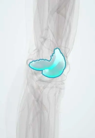
Femoral cartilage !
The femoral cartilage is the smooth layer covering the lower end of the thigh bone, letting the knee glide; it wears down in arthritis.
Official name: Articular cartilage of femur
Opens in the Durgical app, no sign-up needed.
What it is
Articular cartilage: a smooth, slippery layer covering the end of the femur where it meets the tibia and kneecap.
What it does
Lets the joint glide with very little friction and spreads load across the bone.
Key facts
- Where
- Femoral condyles and patellar (trochlear) surface at the knee
- Function
- Hyaline articular cartilage; low-friction gliding; spreads load; avascular, nourished by synovial fluid
Relations
- Below
- Tibial cartilage and the menisci, which cover the outer part of each plateau
- In front
- Patellar cartilage gliding on the trochlea; the suprapatellar pouch above
- Behind
- Posterior condyles, loaded in deep flexion; the capsule and the heads of gastrocnemius beyond
- Deep
- Calcified cartilage layer (tidemark) and the subchondral bone of the condyles
How to find it
With the knee fully bent, the front of the condyles and the edges of the trochlea can be felt on either side of the patellar tendon. At arthroscopy each zone (trochlea, medial and lateral condyles) is inspected and probed for softening or flaps.
Examination
- Wilson test: pain on extending the knee with the tibia internally rotated, relieved by external rotation, suggests osteochondritis dissecans of the medial condyle (low sensitivity)
On imaging
Standing AP and Rosenberg (posteroanterior, knees bent) views show joint-space narrowing better than straight-knee views. MRI cartilage sequences show focal defects and bone marrow oedema; a fluid line behind an osteochondritis fragment marks it as unstable.
Common problems
- Cartilage injuries (focal defects) from sports or a twisting injury.
- Osteoarthritis: gradual wear of the cartilage, causing pain and stiffness.
How it is diagnosed
Symptoms and exam; X-rays show joint-space narrowing in arthritis; MRI shows cartilage defects.
Treatment
- Physical therapy, weight management, activity changes; medicines or injections for pain.
- Surgery for focal defects: microfracture, cell-based repair, or osteochondral grafts (including osteochondral allograft from a donor).
- Knee replacement for advanced arthritis.
Clinical notes
- Juvenile osteochondritis dissecans with open growth plates heals far better than the adult form
- Repair options: microfracture (forms fibrocartilage), osteochondral autograft plugs (mosaicplasty) and cell implantation
- Spontaneous osteonecrosis of the knee affects the weight-bearing medial femoral condyle in older adults, with sudden pain
Development
Articular cartilage is the part of the embryonic hyaline cartilage model that is never replaced by bone. The distal femoral epiphysis begins to ossify just before birth and is the last femoral growth plate to close, in the late teens.
Related conditions
Test yourself
Think of the answer, then tap the question to see it.
Why may an isolated defect of femoral articular cartilage not hurt and heal poorly?
Articular cartilage has no nerves or vessels; pain comes from bone, synovium and capsule.
Where does osteochondritis dissecans classically affect the femur?
The lateral part of the medial femoral condyle, in adolescents.
What tissue does microfracture produce to repair a cartilage defect?
Fibrocartilage, not hyaline cartilage.
Educational information, not medical advice. For any symptom, see a health professional.
Its names
- Official Latin (TA2)
- cartilago articularis TA2 382
- Official Spanish (DeCS)
- Cartílago articular del fémur (cóndilos y tróclea) DeCS D002358
- English (TA2)
- Articular cartilage of femur
Related
Sources: Terminologia Anatomica (TA2); DeCS; Anatomy, Bony Pelvis and Lower Limb, Knee; 8.5 Development of the Appendicular Skeleton (OpenStax (Rice University), 2022) · adapted, CC BY 4.0.
Spotted a mistake?We are always improving for the community. Tell us what is wrong and our team will review it.Report a mistake
Representative model, for educational purposes only.