Nervous system · Head and neck
Choroid plexus !
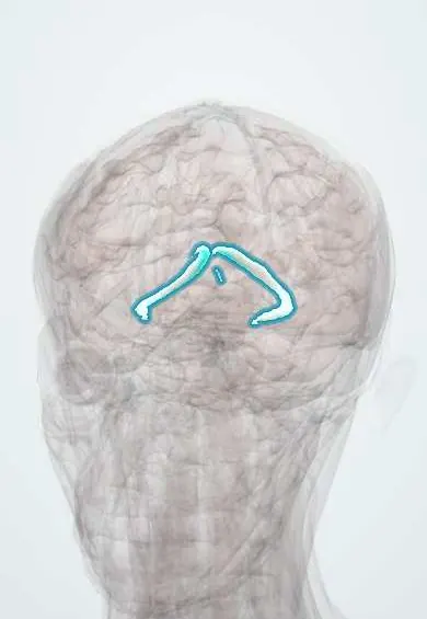
The choroid plexus is made of frond-like tufts of blood vessels inside the brain's ventricles; it produces the cerebrospinal fluid.
Opens in the Durgical app, no sign-up needed.
What it is
Frond-like tufts of vessels inside the ventricles.
What it does
Make the cerebrospinal fluid.
Key facts
- Where
- In the lateral, third and fourth ventricles
- Blood supply
- Anterior choroidal artery (internal carotid) and posterior choroidal arteries (posterior cerebral) for the lateral and third ventricles; PICA for the fourth
- Function
- Makes ~500 mL of CSF per day
Relations
- Course
- A continuous fringe along the choroid fissure, from the interventricular foramen through the body and atrium into the temporal horn; it joins the plexus of the third ventricle roof at the foramina
- Contents
- Fenestrated capillaries in loose connective tissue, covered by a single layer of choroid epithelium (modified ependyma)
- Below
- In the fourth ventricle it hangs from the lower roof and pokes out through the lateral apertures into the cerebellopontine angles
How to find it
The large tuft in the atrium of each lateral ventricle (glomus) is often calcified in adults and shows on CT as a bright spot on both sides, a normal finding. At endoscopy the plexus of the lateral ventricle is followed forward to find the interventricular foramen, through which it continues into the third ventricle.
On imaging
It enhances strongly with contrast because it lies outside the blood–brain barrier. The glomus in the atrium often calcifies in adults, a normal finding on CT.
Clinical notes
- Choroid plexus papilloma, mainly in young children and often in the atrium of the lateral ventricle, can overproduce CSF and cause hydrocephalus
- Choroid plexus cysts are common on prenatal ultrasound and usually vanish; they matter mainly when other anomalies suggest trisomy 18
- Communicating hydrocephalus is a failure of absorption at the arachnoid granulations, not of the plexus
Development
It forms where the ventricle wall stays thin (ependyma lined by pia, the tela choroidea) and tufts of vessels push into the cavity.
Related conditions
Test yourself
Think of the answer, then tap the question to see it.
What forms the blood–CSF barrier in the choroid plexus?
Tight junctions between the choroid epithelial cells covering fenestrated capillaries.
Where is choroid plexus absent in the ventricular system?
In the frontal and occipital horns of the lateral ventricles and in the cerebral aqueduct.
What is the path of CSF from the lateral ventricles to the venous sinuses?
Lateral ventricles, interventricular foramina, third ventricle, aqueduct, fourth ventricle, apertures, subarachnoid space, arachnoid granulations, dural venous sinuses.
Which choroid plexus tumour of young children can cause hydrocephalus by overproducing CSF?
Choroid plexus papilloma, often in the atrium of the lateral ventricle.
Educational information, not medical advice. For any symptom, see a health professional.
Its names
- Official Latin (TA2)
- plexus chorioideus TA2 5409
- Official Spanish (DeCS)
- Plexo Coroideo DeCS D002831
- English (TA2)
- Choroid plexus
Related
Sources: Terminologia Anatomica (TA2); DeCS; Neuroanatomy, Choroid Plexus.
Spotted a mistake?We are always improving for the community. Tell us what is wrong and our team will review it.Report a mistake
Representative model, for educational purposes only.