Nervous system · Head and neck
Third ventricle !
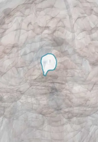
The third ventricle is a narrow fluid-filled cavity in the midline of the brain between the two thalami; it passes cerebrospinal fluid toward the aqueduct.
Opens in the Durgical app, no sign-up needed.
What it is
A narrow midline cavity between the two thalami.
What it does
Passes cerebrospinal fluid from the lateral ventricles to the aqueduct.
Key facts
- Where
- Midline between the thalami
- Blood supply
- Choroid plexus: posterior choroidal arteries
- Function
- CSF -> cerebral aqueduct
Relations
- Lateral
- Thalamus above the hypothalamic sulcus and hypothalamus below; the interthalamic adhesion crosses it in most brains
- Above
- Tela choroidea with its choroid plexus and the internal cerebral veins, then the fornix and the corpus callosum
- In front
- Lamina terminalis, anterior commissure and columns of the fornix; the interventricular foramina at its upper front corners
- Below
- Optic chiasm and hypothalamus, with the infundibulum leading down to the pituitary gland
- Behind
- Pineal gland, habenular and posterior commissures, and the opening of the cerebral aqueduct
How to find it
On a midline sagittal MRI it is the narrow slit between the two thalami. At endoscopy from the frontal horn, the interventricular foramen is found by following the choroid plexus and the thalamostriate and septal veins, which meet at its back edge; through it the mammillary bodies and the thin floor come into view.
On imaging
On sagittal MRI a block at the aqueduct makes the third ventricle balloon, with its floor bowing down and the recesses rounding out, the features checked before a ventriculostomy. A colloid cyst is typically a small, round, often bright (hyperdense) lesion at the foramina on CT.
Clinical notes
- Craniopharyngioma and other suprasellar masses can push up into the floor, causing hydrocephalus plus hypothalamic signs such as great thirst and urine output (diabetes insipidus), weight gain or poor growth
- The transcallosal approach reaches it from above, through the corpus callosum and then the interventricular foramen or the gap between the fornices; damage to both fornices can impair the forming of new memories
- Hypothalamic hamartoma, a mass in its floor or wall, classically causes laughing seizures (gelastic epilepsy) and early puberty
Anatomical variants
- The interthalamic adhesion (massa intermedia) bridges the cavity in most brains but is absent in about 15–20%, more often in men; it is sometimes double; its absence has no known effect
Development
It is the cavity of the diencephalon; the lamina terminalis is the closed front end of the neural tube.
Related conditions
Test yourself
Think of the answer, then tap the question to see it.
What forms the lateral walls of the third ventricle?
The thalamus above the hypothalamic sulcus and the hypothalamus below; the interthalamic adhesion crosses it in most brains.
Why can a colloid cyst of the third ventricle be dangerous?
At the front of the roof it can plug both interventricular foramina: positional headache, sudden hydrocephalus and even sudden death.
Where is the opening made in endoscopic third ventriculostomy?
In the thin floor between the infundibulum and the mammillary bodies, into the cisterns, bypassing an aqueduct block; the basilar artery lies just behind.
Educational information, not medical advice. For any symptom, see a health professional.
Its names
- Official Latin (TA2)
- ventriculus tertius TA2 5769
- Official Spanish (DeCS)
- Tercer Ventrículo DeCS D020542
- English (TA2)
- Third ventricle
Related
Sources: Terminologia Anatomica (TA2); DeCS; Neuroanatomy, Ventricular System; 13.3 Circulation and the Central Nervous System (OpenStax (Rice University), 2022) · adapted, CC BY 4.0; Tratado de anatomía humana (9.ª ed.), tomo II, Libro V, cap. VI: Cerebro (configuración exterior e interior, ventrículos, formaciones coroideas, epífisis).
Spotted a mistake?We are always improving for the community. Tell us what is wrong and our team will review it.Report a mistake
Representative model, for educational purposes only.