Nervous system · Head and neck
Lateral ventricles !
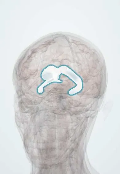
The lateral ventricles are two C-shaped fluid-filled cavities inside the halves of the brain; they hold cerebrospinal fluid and enlarge in hydrocephalus.
Official name: Lateral ventricle
Opens in the Durgical app, no sign-up needed.
What it is
Two C-shaped fluid cavities inside the hemispheres.
What it does
Hold cerebrospinal fluid; they enlarge in hydrocephalus.
Key facts
- Where
- Inside each hemisphere (frontal, occipital, temporal horns)
- Blood supply
- Choroid plexus: anterior and posterior choroidal arteries
- Function
- CSF -> interventricular foramina -> third ventricle
Relations
- Above
- Corpus callosum, forming the roof of the frontal horn and body (and, as the tapetum, the roof and lateral wall of the occipital and temporal horns); in the roof of the temporal horn, the tail of the caudate nucleus and the stria terminalis
- Medial
- Septum pellucidum between the two frontal horns and bodies; the fornix along the lower medial wall of the body; on the medial wall of the occipital horn, the bulb (forceps major) above and the calcar avis (raised by the calcarine sulcus) below
- Lateral
- Head of the caudate nucleus bulging into the frontal horn; the optic radiation outside the temporal and occipital horns
- Below
- Floor of the body, lateral to medial: body of the caudate, the groove with the stria terminalis and thalamostriate vein, the thalamus, the choroid plexus and the fornix; floor of the temporal horn: the hippocampus with the fimbria, and the collateral eminence laterally
- Contents
- CSF, and the choroid plexus in the body, atrium and temporal horn
How to find it
Kocher point, the usual entry for a frontal horn catheter, lies about 11 cm behind the nasion and 3 cm from the midline (near the mid-pupillary line), in front of the coronal suture; the catheter is aimed toward the inner corner of the eye on that side and the front of the ear (some texts: 10–11 cm and 2.5–3 cm).
On imaging
Cranial ultrasound through the anterior fontanelle in babies; CT or MRI later. The Evans index (widest span of the frontal horns divided by the widest inner skull diameter) above 0.3 indicates enlarged ventricles. A calcified choroid plexus in the atrium is a common normal finding.
Common problems
- Hydrocephalus: fluid builds up and the ventricles enlarge, raising pressure (headache, vomiting; in babies a growing head).
How it is diagnosed
Ultrasound in babies; CT or MRI.
Treatment
- A shunt (a thin tube from the ventricle to the abdomen) or an endoscopic opening (third ventriculostomy).
Clinical notes
- Ventriculoperitoneal shunt: a catheter in the frontal or occipital horn drains CSF through a valve under the skin into the abdomen; blockage, infection and overdrainage are its problems
- External ventricular drain: placed through Kocher point into the frontal horn in emergencies
- Preterm babies bleed from the germinal matrix next to the caudate head, and the blood can fill the ventricles
- In normal-pressure hydrocephalus, improved walking after removing CSF by lumbar puncture supports a shunt
Anatomical variants
- Mild asymmetry between the two lateral ventricles is common and usually of no significance
- Cavum septi pellucidi: a fluid space between the two leaves of the septum pellucidum; normal in the fetus and newborn, it persists into adult life in some people, at times extending backward as a cavum vergae
- The occipital horns vary most in size and are often asymmetric, the left usually the larger; the calcar avis is occasionally absent
Development
Each one is the cavity of a side pouch of the telencephalon, a remnant of the hollow neural tube. As the hemisphere grows forward, up, back and then down around the insula, the cavity is drawn into its C shape, ending in the temporal horn.
Memory aid
Normal-pressure hydrocephalus: 'wet, wobbly and wacky' (incontinence, gait, dementia).
Related conditions
- Fumarase deficiency
- Neurocysticercosis
- Hydrocephalus
- Zika virus infection
- Cryptococcal meningitis
- Raised intracranial pressure and brain herniation
- Tuberous sclerosis complex
- Prematurity and its complications
- Congenital (TORCH) infections: syphilis, toxoplasmosis, CMV and Zika
- Intraventricular haemorrhage of prematurity
Test yourself
Think of the answer, then tap the question to see it.
What are the parts of each lateral ventricle, and how does it drain?
Frontal horn, body, atrium, occipital horn and temporal horn; it drains into the third ventricle through an interventricular foramen (of Monro).
Why are ventricular catheters aimed at the frontal horn of the lateral ventricle?
The frontal and occipital horns have no choroid plexus; the frontal horn is reached through Kocher point.
What is the triad of normal-pressure hydrocephalus?
Gait trouble, urinary incontinence and dementia with large ventricles; described by the Colombian neurosurgeon Salomón Hakim (Hakim–Adams syndrome).
What value of the Evans index indicates enlarged lateral ventricles?
Above 0.3: the widest span of the frontal horns divided by the widest inner skull diameter.
Educational information, not medical advice. For any symptom, see a health professional.
Its names
- Official Latin (TA2)
- ventriculus lateralis TA2 5639
- Official Spanish (DeCS)
- Ventrículos Laterales DeCS D020547
- English (TA2)
- Lateral ventricle
Related
Sources: Terminologia Anatomica (TA2); DeCS; Neuroanatomy, Ventricular System; Anatomy of the Human Body (20th ed.): Development of the Nervous System (Lea & Febiger (Gray's Anatomy, 1918), 1918) · adapted, Public domain; 13.1 The Embryologic Perspective (OpenStax (Rice University), 2022) · adapted, CC BY 4.0; Tratado de anatomía humana (9.ª ed.), tomo II, Libro V, cap. VI: Cerebro (configuración exterior e interior, ventrículos, formaciones coroideas, epífisis).
Spotted a mistake?We are always improving for the community. Tell us what is wrong and our team will review it.Report a mistake
Representative model, for educational purposes only.