Hydrocephalus !
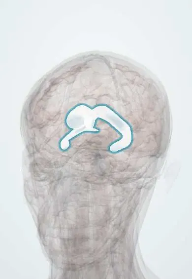
Hydrocephalus is a build-up of cerebrospinal fluid in the brain's cavities, which widen and can raise pressure inside the skull.
Updated 2026-10-29
Structures involved
Tap one to see what it is and open it in 3D.
- Lateral ventricles
- Third ventricle
- Cerebral aqueduct
- Fourth ventricle
- Choroid plexus
- Superior sagittal sinus
- Cerebellar tonsils
- Corpus callosum
Microorganisms that cause it
In histology
- Gray and white matter, cerebellar cortex, meninges
- Blood–brain barrier, choroid plexus and cerebrospinal fluid
In embryology
- Neurulation and closure of the neural tube
- Skull: neurocranium, viscerocranium, sutures and fontanelles
- Craniosynostosis
- Achondroplasia (FGFR3)
- Brain vesicles: from three to five and what each becomes
- Ventricles and cerebrospinal fluid
- Neural tube defects: anencephaly, spina bifida and encephalocele
- Chiari malformations
- Congenital hydrocephalus: aqueductal stenosis
- Dandy–Walker malformation
In the clinical exam
In physiology
What it is
Abnormal build-up of cerebrospinal fluid in the ventricles of the brain, which enlarge and may raise intracranial pressure. It can be congenital or acquired, and obstructive or communicating.
Who gets it
Occurs from infancy to old age: congenital forms and those linked to prematurity and neural tube defects in children, and normal pressure hydrocephalus in older adults. In Colombia, neurocysticercosis and infections are important acquired causes.
Causes
- Obstruction of flow: aqueductal stenosis, tumours (especially of the posterior fossa), cysts such as ventricular neurocysticercosis
- Impaired absorption: after subarachnoid haemorrhage or meningitis
- Congenital malformations: Chiari II with myelomeningocele, Dandy-Walker malformation
- Intraventricular haemorrhage in premature infants
- Rarely overproduction (choroid plexus papilloma)
Risk factors
- Prematurity
- Neural tube defects
- Meningitis and neurocysticercosis
- Subarachnoid or intraventricular haemorrhage
- Brain tumours and head injury
- Older age (normal pressure hydrocephalus)
Pathophysiology
CSF made by the choroid plexus flows from the lateral ventricles through the third ventricle, aqueduct and fourth ventricle to the subarachnoid space, then drains to the venous sinuses. Blocked flow within the ventricles causes obstructive hydrocephalus; impaired absorption causes communicating hydrocephalus. In infants with open sutures the head enlarges; in older patients pressure rises quickly.
Classification
- Obstructive (non-communicating)
- Communicating
- Normal pressure hydrocephalus (older adults)
- Congenital or acquired
- Acute or chronic
Symptoms and signs
- Infants: rapidly growing head circumference, bulging fontanelle, separated sutures, irritability, vomiting, sunset eyes
- Older children and adults: headache worse in the morning, nausea, vomiting, drowsiness, papilloedema
- Upward gaze palsy (Parinaud syndrome) with dorsal midbrain compression
- Normal pressure hydrocephalus: gait disturbance, urinary incontinence and cognitive decline
- Shunt malfunction: return of headache, vomiting and drowsiness
Diagnosis
- Clinical
- Head circumference charts and fontanelle examination in infants; symptoms of raised pressure and fundoscopy in older patients; the gait-incontinence-dementia triad in normal pressure hydrocephalus.
- Tests
- CSF studies when infection is suspected. In normal pressure hydrocephalus, a high-volume lumbar tap test or lumbar drainage to see whether gait improves.
- Imaging
- Cranial ultrasound through the fontanelle in infants. CT or MRI shows enlarged ventricles and the site of obstruction; MRI is better for aqueduct and posterior fossa lesions.
Differential diagnosis
- Brain atrophy with ex vacuo ventricular enlargement
- Benign enlargement of subarachnoid spaces in infancy
- Idiopathic intracranial hypertension
- Alzheimer disease and Parkinson disease (versus normal pressure hydrocephalus)
- Brain tumour or abscess
Goal of treatment
Divert or restore CSF flow to relieve raised pressure and protect brain development and function, and treat the cause.
Treatment
- Acute
- Acute obstructive hydrocephalus with deterioration: emergency external ventricular drain.
- Surgical
- Ventricular shunt, usually ventriculoperitoneal, with a valve that controls drainage.
- Surgical
- Endoscopic third ventriculostomy for selected obstructive cases, avoiding a permanent shunt.
- Surgical
- Removal of the cause when possible: tumour resection, endoscopic removal of ventricular cysticerci.
- Follow-up
- Lifelong follow-up of shunted patients for malfunction or infection; developmental follow-up in children.
Complications
- Shunt obstruction, infection or over-drainage
- Brain herniation and death if untreated
- Visual loss from chronic papilloedema
- Developmental delay, learning difficulties and epilepsy
- Subdural collections after drainage
Prognosis
Depends on the cause, age and speed of treatment. Many children reach good developmental outcomes, although shunt revisions are common. In normal pressure hydrocephalus, gait responds best to shunting.
Prevention
- Folic acid before conception to reduce neural tube defects
- Prenatal care to reduce prematurity
- Prevention and treatment of neurocysticercosis and meningitis
- Vaccination against meningitis-causing bacteria
Test yourself
Think of the answer, then tap the question to see it.
What is the difference between obstructive and communicating hydrocephalus?
Obstructive: flow is blocked within the ventricular system; communicating: CSF reaches the subarachnoid space but is poorly absorbed.
What is the triad of normal pressure hydrocephalus?
Gait disturbance, urinary incontinence and cognitive decline.
Why does the head enlarge in infants with hydrocephalus?
Their cranial sutures and fontanelles are still open, so the skull expands with the raised pressure.
Which parasitic infection is an important cause of hydrocephalus in Colombia?
Neurocysticercosis, through ventricular cysts or arachnoiditis.
What is the most common surgical treatment for hydrocephalus?
A ventriculoperitoneal shunt; endoscopic third ventriculostomy is an option in selected obstructive cases.
Related conditions
- Neurocysticercosis
- Bacterial meningitis
- Subarachnoid haemorrhage
- Spina bifida (myelomeningocele)
- Glioblastoma
- Alzheimer disease
- Vestibular schwannoma (acoustic neuroma)
Educational information, not medical advice. For any symptom, see a health professional.
Sources: Hydrocephalus (StatPearls (NCBI Bookshelf), 2023).
Spotted a mistake?We are always improving for the community. Tell us what is wrong and our team will review it.Report a mistake
Representative model, for educational purposes only.