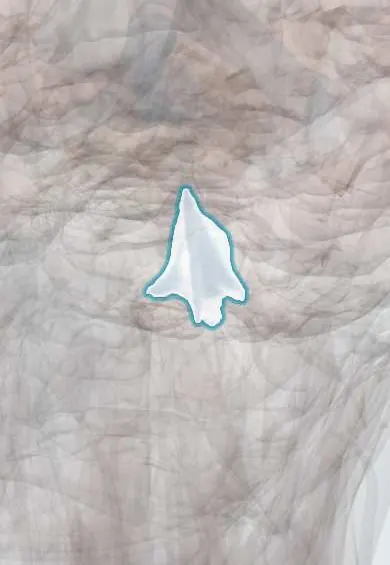
Nervous system · Head and neck
Fourth ventricle !
The fourth ventricle is a tent-shaped cavity between the brainstem and the cerebellum; it lets cerebrospinal fluid out around the brain and spinal cord.
Opens in the Durgical app, no sign-up needed.
What it is
A tent-shaped cavity between the brainstem and the cerebellum.
What it does
Lets cerebrospinal fluid out around the brain and spinal cord.
Key facts
- Where
- Between the pons/medulla and the cerebellum
- Blood supply
- Choroid plexus: PICA
- Function
- CSF exits via the median and lateral apertures to the subarachnoid space
Relations
- In front
- Floor formed by the pons above and the medulla oblongata below
- Behind
- Cerebellum; roof formed above by the superior medullary velum, stretched between the superior cerebellar peduncles (the trochlear nerves cross inside it and emerge beside its frenulum, below the inferior colliculi), and below by the inferior medullary velum and tela choroidea with its choroid plexus
- Lateral
- Cerebellar peduncles; lateral recesses curving round the inferior peduncles to the lateral apertures (Luschka), which open into the cerebellopontine angle beside the flocculus, just behind the glossopharyngeal and vagus rootlets and below the facial and vestibulocochlear nerves, with a tuft of choroid plexus poking through
- Above
- Cerebral aqueduct
- Below
- Obex at the lower tip of the floor (calamus scriptorius), where it narrows into the central canal of the medulla and spinal cord; the median aperture (Magendie) opens in the lower roof just above it, into the cisterna magna
How to find it
On a midline sagittal MRI it is the tent-shaped space with the pons and medulla in front and the cerebellum behind, its peak (fastigium) pointing back into the cerebellum. Surgeons reach it through a midline suboccipital craniotomy, lifting the tonsils to enter from below.
On imaging
Sagittal MRI shows a tent whose peak (fastigium) points into the cerebellum. A normal fourth with large lateral and third ventricles puts the block at the aqueduct; in Dandy–Walker malformation it balloons into a posterior fossa cyst. Neurocysticercal cysts may show only a thin wall and need thin-slice MRI.
Clinical notes
- Surgeons reach it from below through the cerebellomedullary fissure (telovelar approach), avoiding splitting the vermis; injury to the facial colliculus is a key risk
- Ventricular cysticerci are often removed endoscopically, as they block flow and do not resolve readily
Anatomical variants
- The lateral apertures are occasionally absent or closed (a few percent in classic dissection series)
Development
It is the cavity of the hindbrain. Its roof stays thin, carries the choroid plexus and later opens through the median and lateral apertures; in Dandy–Walker malformation the roof balloons into a cyst and the vermis is small.
Memory aid
Magendie = Median aperture; Luschka = Lateral apertures.
Related conditions
- Neurocysticercosis
- Hydrocephalus
- Childhood brain tumours (medulloblastoma, pilocytic astrocytoma, craniopharyngioma)
Test yourself
Think of the answer, then tap the question to see it.
Through which openings does CSF leave the fourth ventricle?
The median aperture (Magendie) and the two lateral apertures (Luschka), into the subarachnoid space.
Which parasitic disease, common in Colombia, can block the fourth ventricle?
Neurocysticercosis: larval cysts of the pork tapeworm lodge in the ventricles, most often the fourth, causing obstructive hydrocephalus.
Which childhood tumours fill the fourth ventricle?
Medulloblastoma from the roof, ependymoma from the floor squeezing out through the apertures, and pilocytic astrocytoma.
What forms the floor of the fourth ventricle?
The pons above and the medulla oblongata below (rhomboid fossa), with landmarks such as the facial colliculus and the hypoglossal and vagal trigones.
Educational information, not medical advice. For any symptom, see a health professional.
Its names
- Official Latin (TA2)
- ventriculus quartus TA2 5966
- Official Spanish (DeCS)
- Cuarto Ventrículo DeCS D020546
- English (TA2)
- Fourth ventricle
Related
Sources: Terminologia Anatomica (TA2); DeCS; Neuroanatomy, Fourth Ventricle; Anatomy of the Human Body (20th ed.): Development of the Nervous System (Lea & Febiger (Gray's Anatomy, 1918), 1918) · adapted, Public domain; 13.1 The Embryologic Perspective (OpenStax (Rice University), 2022) · adapted, CC BY 4.0; Tratado de anatomía humana (9.ª ed.), tomo II, Libro V, sección 2 (Encéfalo), capítulos I-V: bulbo, protuberancia, cerebelo, cuarto ventrículo, mesencéfalo.
Spotted a mistake?We are always improving for the community. Tell us what is wrong and our team will review it.Report a mistake
Representative model, for educational purposes only.