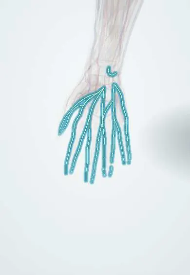
Digital nerves !
The digital nerves run along both sides of each finger on the palm side; they give the fingertips their fine sense of touch.
Official name: Palmar digital nerves
Opens in the Durgical app, no sign-up needed.
What it is
Thin nerves along each side of every finger.
What it does
Give the fingertips their fine touch.
Key facts
- Roots
- Common palmar digital branches of the median and ulnar nerves
- Moves
- Median ones also supply lumbricals 1–2
- Feeling from
- Palmar skin of the fingers and their nail beds
Relations
- Course
- Common palmar digital nerves run down the palm under the superficial palmar arch and palmar aponeurosis, between the flexor tendons, and split near the webs into proper digital nerves that run along each side of the finger to the pulp and nail bed
- In front
- In the finger, Grayson's ligaments and the palmar skin
- Behind
- The digital artery, dorsal to the nerve in the finger, and Cleland's ligaments
- Deep
- The flexor tendon sheath and its pulleys, along whose sides the nerves run
How to find it
The neurovascular bundles run along each side of the finger, palmar to the midaxial line (the line joining the ends of the finger creases); this is why midaxial incisions stay dorsal to them.
Examination
- Static two-point discrimination on the radial and ulnar side of each pulp: normal fingertips separate two points a few millimetres apart (usually under about 6 mm)
- Wrinkle test: after soaking in warm water, skin that has lost its nerve supply stays smooth; useful in young children
- A Tinel's sign that moves down the finger after repair tracks the regenerating nerve
On imaging
Not seen on radiographs. High-frequency ultrasound follows the common and proper palmar digital nerves next to their arteries, whose Doppler flow is the landmark, and can show a cut nerve end or a neuroma after a laceration. Most acute injuries are diagnosed by testing sensation and explored at surgery; radiographs look for a foreign body or an associated fracture.
Clinical notes
- Glass and knife cuts often divide digital nerves; repair under magnification, sometimes with a nerve graft or conduit, restores protective feeling
- A digital nerve block (at the base of the finger or through the flexor sheath) numbs the finger for wound repair and nail procedures
- Dupuytren's disease: a spiral cord can pull the neurovascular bundle toward the midline and the surface, where it is at risk during fasciectomy
- Bowler's thumb: scarring around the ulnar digital nerve of the thumb from repeated pressure of the ball's thumb hole
Anatomical variants
- Connections between the median and ulnar digital branches in the palm are frequent, so the whole ring finger may be supplied by one nerve and the border between territories shifts
Development
Their sensory cell bodies lie in dorsal root ganglia, which come from the neural crest. The fingers part in weeks 6–7 by cell death between the rays; in syndactyly the common digital nerve and artery often divide further toward the tip, which limits how far the web can be released.
Related conditions
Test yourself
Think of the answer, then tap the question to see it.
In the finger, where does the proper digital nerve lie relative to the digital artery?
Palmar to the artery; in the palm, the common digital nerves lie deep to the superficial palmar arch.
What is checked after a finger cut before any local anaesthetic, and why?
Two-point discrimination on each side of the pulp; loss on one side means that digital nerve is probably divided.
What does the wrinkle test show, and in whom is it useful?
After soaking in warm water, skin that has lost its digital nerve supply stays smooth; useful in young children.
Educational information, not medical advice. For any symptom, see a health professional.
Its names
- Official Latin (TA2)
- rami digitales palmares communes nervi mediani TA2 6465
- Official Spanish (DeCS)
- Ramos digitales palmares comunes y propios de los nervios mediano y ulnar
- English (TA2)
- Palmar digital nerves
Related
Sources: Terminologia Anatomica (TA2); Anatomy, Shoulder and Upper Limb, Hand Cutaneous Innervation; 13.1 The Embryologic Perspective (OpenStax (Rice University), 2022) · adapted, CC BY 4.0; 8.5 Development of the Appendicular Skeleton (OpenStax (Rice University), 2022) · adapted, CC BY 4.0.
Spotted a mistake?We are always improving for the community. Tell us what is wrong and our team will review it.Report a mistake
Representative model, for educational purposes only.