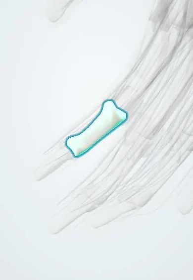
Middle phalanx, index finger !
The middle phalanx of the index finger is its middle bone, between the two finger joints; tendons that bend and straighten the finger attach here.
Official name: Middle phalanx of the index finger
Opens in the Durgical app, no sign-up needed.
What it is
The middle bone of the index finger.
What it does
One of the finger bones; the joints between them let the finger curl.
Key facts
- Joints with
- Proximal phalanx (PIP), distal phalanx (DIP)
- Landmarks
- Base, shaft, head; FDS inserts on its sides
Relations
- Above
- Head of the proximal phalanx at the PIP joint, with the volar plates (fingers) and finger collateral ligaments
- In front
- FDP tendon on its way to the distal phalanx, between the FDS slips
- Behind
- Central slip at the base; lateral bands passing on each side toward the distal phalanx
How to find it
The short bony segment on the back of the index between the two finger joints; the middle finger crease on the palm marks the PIP joint at its base.
Examination
- Superficialis test: with the other fingers held straight, the person bends only the index at the PIP joint; failure suggests an FDS tendon cut (the index FDS can sometimes be helped by its own FDP, so compare both sides)
- Elson test: with the PIP bent 90° over a table edge, weak middle-phalanx extension and a rigid DIP suggest a central slip rupture
On imaging
A true lateral view of the index is the key film: it shows a palmar base fragment from the volar plate and any dorsal subluxation of the base (a gap shaped like a V on the back of the joint).
Clinical notes
- Central slip rupture at its base leads to a boutonnière deformity (PIP flexed, DIP hyperextended)
- In a dorsal PIP fracture-dislocation, the loss of a large part of its palmar base lets the phalanx slide backward; a congruent joint on the true lateral view is the aim of treatment
Development
Like the other phalanges, it ossifies from a shaft centre before birth and one secondary centre at its base; middle phalanges are generally the last phalanges to start ossifying.
Test yourself
Think of the answer, then tap the question to see it.
Which tendons insert on the middle phalanx of the index finger?
The two slips of flexor digitorum superficialis on the sides and the extensor central slip on the dorsal base.
What deformity follows rupture of the central slip at the base of the middle phalanx?
Boutonnière deformity: PIP flexed and DIP hyperextended.
What injury causes a volar plate avulsion fragment at the palmar base of the middle phalanx?
Hyperextension of the PIP joint; a large fragment can let the joint sublux dorsally.
Educational information, not medical advice. For any symptom, see a health professional.
Its names
- Official Latin (TA2)
- phalanx media manus TA2 1278
- Official Spanish (DeCS)
- Falange media del índice
- English (TA2)
- Middle phalanx of the index finger
Related
- Extensor tendons
- FDP
- FDP tendons
- FDS
- FDS tendons
- Finger joint capsules
- Finger pulleys
- Flexor tendon sheaths
Sources: Terminologia Anatomica (TA2); Anatomy, Shoulder and Upper Limb, Hand Bones; 8.2 Bones of the Upper Limb (OpenStax (Rice University), 2022) · adapted, CC BY 4.0; 8.5 Development of the Appendicular Skeleton (OpenStax (Rice University), 2022) · adapted, CC BY 4.0; Anatomy of the Human Body (20th ed.): Articulations of the Digits (Lea & Febiger (Gray's Anatomy, 1918), 1918) · adapted, Public domain.
Spotted a mistake?We are always improving for the community. Tell us what is wrong and our team will review it.Report a mistake
Representative model, for educational purposes only.