Nervous system · Head and neck
Falx cerebri !
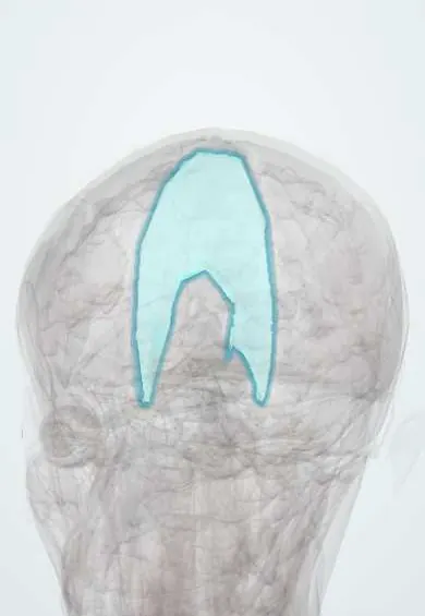
The falx cerebri is a sickle-shaped fold of dura between the two halves of the brain; it holds the brain steady.
Opens in the Durgical app, no sign-up needed.
What it is
A sickle-shaped fold of dura between the two hemispheres.
What it does
Holds the brain steady; brain can shift under it (subfalcine herniation).
Key facts
- Attaches
- Crista galli in front to the internal occipital protuberance behind, joining the tentorium
- Function
- Dural fold between the hemispheres; holds the superior and inferior sagittal sinuses; subfalcine herniation passes under it
Relations
- Contents
- Superior sagittal sinus in its attached upper border, inferior sagittal sinus in its free lower border, straight sinus along its junction with the tentorium cerebelli
- Lateral
- Medial surfaces of the two cerebral hemispheres, in the longitudinal fissure
- Below
- Corpus callosum below its free edge, with the cingulate gyrus and the anterior cerebral artery branches between edge and callosum
How to find it
On axial CT or MRI it is the thin midline line between the hemispheres, the reference for measuring midline shift. At surgery it lies at the bottom of the interhemispheric fissure: its fixed upper edge holds the superior sagittal sinus and its free lower edge the inferior sagittal sinus, just above the corpus callosum.
On imaging
On axial CT or MRI it is a thin midline line whose displacement measures midline shift. Blood along it appears as a thick, bright falx (interhemispheric subdural haematoma); calcified plaques are a common normal finding.
Clinical notes
- Falcine meningiomas grow from it, often into both sides, and may involve the sagittal sinuses
- Interhemispheric surgical approaches work along it to reach the corpus callosum and the midline
Development
In holoprosencephaly the forebrain fails to divide into two hemispheres and the falx is absent or only partly formed.
Related conditions
Test yourself
Think of the answer, then tap the question to see it.
Where does the falx cerebri attach?
From the crista galli in front to the internal occipital protuberance behind, where it joins the tentorium.
Which venous sinuses run in the falx cerebri?
Superior sagittal sinus in its attached upper border, inferior sagittal sinus in its free lower border, straight sinus along its junction with the tentorium.
In subfalcine herniation, which artery can be pinched against the falx cerebri and with what result?
The anterior cerebral artery, causing an infarct with weakness of the opposite leg.
Educational information, not medical advice. For any symptom, see a health professional.
Its names
- Official Latin (TA2)
- falx cerebri TA2 5374
- Official Spanish (DeCS)
- Hoz del cerebro
- English (TA2)
- Falx cerebri
- Also called
- cerebral falx
Related
Sources: Terminologia Anatomica (TA2); Neuroanatomy, Falx Cerebri.
Spotted a mistake?We are always improving for the community. Tell us what is wrong and our team will review it.Report a mistake
Representative model, for educational purposes only.