Nervous system · Head and neck
Tentorium !
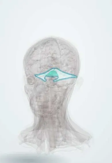
The tentorium cerebelli is a tent-like sheet of dura that separates the cerebellum from the cerebrum above; brain can herniate through its opening.
Official name: Tentorium cerebelli
Opens in the Durgical app, no sign-up needed.
What it is
A tent of dura between the cerebrum and the cerebellum.
What it does
Its notch is where the temporal lobe can herniate and press on nerve III.
Key facts
- Attaches
- Petrous ridges, clinoid processes and the grooves of the transverse sinuses
- Function
- Dural roof over the cerebellum; its free edge (tentorial notch) surrounds the midbrain; uncal herniation here compresses III
Relations
- Contents
- Straight sinus along its junction with the falx cerebri, the transverse sinuses in its attached outer border, the superior petrosal sinuses along the petrous ridges
- Above
- Occipital lobe and the lower surface of the temporal lobe
- Below
- Upper surface of the cerebellum
- Medial
- Midbrain in the tentorial notch; the oculomotor nerve passes forward between the posterior cerebral artery and the superior cerebellar artery, and the trochlear nerve runs along the free edge
How to find it
On coronal and sagittal MRI it is the tent-shaped sheet over the cerebellum, bright after contrast. Its outer edge follows the grooves of the transverse sinuses and the upper border of the petrous bone; its free edge forms the tentorial notch around the midbrain, where the oculomotor and trochlear nerves run close to it.
On imaging
Coronal and sagittal MRI show a thin, enhancing tent-shaped sheet; tentorial subdural blood layers along it. On CT, effaced cisterns around the midbrain suggest transtentorial herniation.
Clinical notes
- Compression of the posterior cerebral artery in uncal herniation can cause an occipital infarct with loss of the opposite visual field
- Downward displacement of the brainstem through the notch can tear small perforating vessels: Duret haemorrhages in the midbrain and pons
- Posterior fossa masses can push the cerebellum upward through the notch or drive the cerebellar tonsils down into the foramen magnum
Related conditions
Test yourself
Think of the answer, then tap the question to see it.
Which compartments does the tentorium cerebelli divide the cranial cavity into?
Supratentorial (cerebral hemispheres) and infratentorial (posterior fossa: cerebellum, pons, medulla).
What does uncal herniation over the tentorium compress?
The oculomotor nerve (dilated pupil, then ptosis, eye down and out) and the posterior cerebral artery.
Why can compression of the posterior cerebral artery in uncal herniation cause visual loss?
It can cause an occipital infarct with loss of the opposite visual field.
Educational information, not medical advice. For any symptom, see a health professional.
Its names
- Official Latin (TA2)
- tentorium cerebelli TA2 5375
- Official Spanish (DeCS)
- Tienda del cerebelo
- English (TA2)
- Tentorium cerebelli
- Also called
- cerebellar tentorium
Related
Sources: Terminologia Anatomica (TA2); Neuroanatomy, Tentorium Cerebelli.
Spotted a mistake?We are always improving for the community. Tell us what is wrong and our team will review it.Report a mistake
Representative model, for educational purposes only.