Cardiovascular system · Head and neck
Superior sagittal sinus !
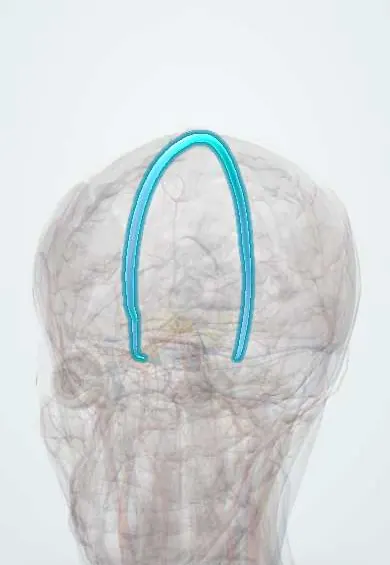
The superior sagittal sinus is a large vein channel along the top midline of the skull; it drains the brain surface and returns spinal fluid to the blood.
Opens in the Durgical app, no sign-up needed.
What it is
A large vein channel in the top edge of the falx, along the midline of the skull.
What it does
Drains the brain's surface and reabsorbs cerebrospinal fluid; it can clot (venous sinus thrombosis).
Key facts
- Drains
- Superficial cerebral veins; absorbs CSF via arachnoid granulations
- Into
- Confluence of sinuses
Relations
- Course
- Begins near the crista galli, runs back in the attached upper border of the falx cerebri along the midline of the skull vault, widening as it goes, and ends at the internal occipital protuberance in the confluence of sinuses
- Above
- Inner table of the skull in the midline; emissary veins through the parietal bones link it to the scalp veins, a route for scalp infection to spread inward
- Below
- The falx cerebri, hanging between the two hemispheres
- Lateral
- Lateral venous lacunae with arachnoid granulations, and the superior cerebral veins entering it
How to find it
Its line follows the midline of the scalp from the root of the nose to the external occipital protuberance, under the sagittal suture. In infants it lies just under the anterior fontanelle; burr holes and craniotomies are kept a little off the midline to avoid it.
On imaging
CT or MR venography shows it as a midline channel. In thrombosis, non-contrast CT may show a bright clot (dense triangle); contrast CT shows the empty delta sign, a dark triangular clot ringed by enhancing dura in the back of the sinus.
Clinical notes
- Venous infarcts from its thrombosis are often haemorrhagic and do not fit an arterial territory; bilateral parasagittal lesions suggest this sinus
- Midline skull fractures and surgery near it can cause heavy bleeding; its front third can be tied off with less risk than the back two thirds, whose closure causes venous infarction
- Parasagittal and falcine meningiomas may invade it, which limits how much tumour can be removed
Anatomical variants
- At the confluence it usually turns mainly into the right transverse sinus, while the straight sinus favours the left (some texts describe a fully shared confluence)
Development
It grows from a plexus of small veins in the midline that extends forward and then backward with the cerebral hemispheres; its front part is completed first, and the back part forms as the hemispheres enlarge.
Related conditions
- Cerebral venous sinus thrombosis
- Traumatic brain injury
- Bacterial meningitis
- Subdural haematoma
- Hydrocephalus
Test yourself
Think of the answer, then tap the question to see it.
What is the drainage route from the superior sagittal sinus to the internal jugular vein?
Superior sagittal sinus, confluence of sinuses, transverse sinus, sigmoid sinus, internal jugular vein.
Why does a blocked superior sagittal sinus raise intracranial pressure?
Arachnoid granulations push into it and return most CSF to the blood.
What are the features of superior sagittal sinus thrombosis?
Headache, papilloedema, seizures and focal deficits, often with haemorrhagic venous infarcts that do not fit an arterial territory.
What does tearing of the bridging veins that drain into the superior sagittal sinus cause?
A subdural haematoma: the superior cerebral veins cross the subdural space to reach the sinus.
Educational information, not medical advice. For any symptom, see a health professional.
Its names
- Official Latin (TA2)
- sinus sagittalis superior TA2 4856
- Official Spanish (DeCS)
- Seno Sagital Superior DeCS D054063
- English (TA2)
- Superior sagittal sinus
- Also called
- Seno longitudinal superior
Related
Sources: Terminologia Anatomica (TA2); DeCS; Neuroanatomy, Superior Sagittal Sinus; Neuroanatomy, Dural Venous Sinuses; Anatomy of the Human Body (20th ed.): Development of the Vascular System (Lea & Febiger (Gray's Anatomy, 1918), 1918) · adapted, Public domain.
Spotted a mistake?We are always improving for the community. Tell us what is wrong and our team will review it.Report a mistake
Representative model, for educational purposes only.