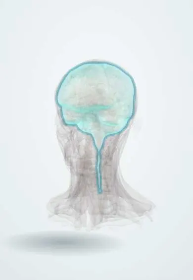
Subdural haematoma !
A subdural haematoma is blood collecting under the brain's outer covering from torn veins, after major trauma or, in older adults, minor falls.
Updated 2026-10-29
Structures involved
Tap one to see what it is and open it in 3D.
In the clinical exam
In physiology
Why it happens
- Hemorrhage: petechiae, purpura, ecchymosis, hematoma, and what blood loss does
- Mechanical and electrical injury: abrasion, contusion, laceration, wounds
What it is
A collection of blood between the dura mater and the arachnoid, usually from torn bridging veins. It can be acute after major trauma or chronic in older adults after minor or forgotten injuries.
Who gets it
Acute forms follow high-energy trauma at any age. Chronic forms mainly affect older adults, especially those on anticoagulant or antiplatelet drugs or with alcohol use disorder; their frequency rises as populations age.
Causes
- Tearing of bridging veins that cross from the cortex to the dural venous sinuses
- Acceleration-deceleration head injury (falls, road traffic accidents)
- Minor head injury in older adults (chronic form)
- Non-accidental head injury in infants (shaking)
Risk factors
- Older age (brain atrophy stretches bridging veins)
- Anticoagulant or antiplatelet therapy
- Alcohol use disorder
- Falls and dementia
- Coagulopathy, thrombocytopenia, dialysis
- Infancy (abusive head trauma)
Pathophysiology
Bridging veins tear and bleed into the potential space beneath the dura, spreading over the hemisphere in a crescent. In acute cases the clot and underlying brain injury raise intracranial pressure quickly. In chronic cases a slow bleed is encapsulated by membranes with fragile new vessels that rebleed, so the collection enlarges over weeks.
Classification
- Acute (hours to a few days; hyperdense on CT)
- Subacute (days to about two weeks; may be isodense)
- Chronic (weeks; hypodense on CT)
- Acute-on-chronic (mixed density)
Symptoms and signs
- Acute: reduced consciousness soon after a severe head injury
- Chronic: headache that is gradual and worsening
- Confusion, memory loss, personality change, drowsiness (may mimic dementia)
- Unilateral weakness, gait disturbance, falls
- Speech disturbance, seizures
Diagnosis
- Clinical
- History of trauma (often minor or absent in chronic cases), anticoagulant use and a fluctuating or progressive neurological picture; serial Glasgow Coma Scale scoring.
- Tests
- Coagulation studies, platelet count and full blood count; renal function. Skeletal survey and ophthalmological examination in infants with suspected abuse.
- Imaging
- Non-contrast head CT: a crescent-shaped collection that crosses suture lines but not the falx or tentorium. Density changes from hyperdense to hypodense with age. MRI helps with subacute isodense collections.
Differential diagnosis
- Epidural haematoma
- Dementia or delirium of other cause
- Ischaemic stroke
- Brain tumour
- Subdural hygroma or empyema
Goal of treatment
Relieve compression of the brain, stop further bleeding and prevent recurrence, while correcting coagulation problems.
Treatment
- Acute
- Resuscitation and reversal of anticoagulant or antiplatelet effects; control of intracranial pressure.
- Surgical
- Acute haematomas with mass effect or neurological decline: urgent craniotomy and evacuation, sometimes decompressive craniectomy.
- Surgical
- Symptomatic chronic haematomas: drainage through burr holes, often with a temporary drain to reduce recurrence.
- Procedure
- Middle meningeal artery embolisation, used in some centres for recurrent or selected chronic haematomas.
- Follow-up
- Small collections with few symptoms: observation with serial examination and repeat CT.
Complications
- Brain herniation and death (acute form)
- Recurrence of chronic haematoma
- Seizures
- Persistent cognitive or motor deficits
- Complications of stopping antithrombotic treatment
Prognosis
Acute subdural haematoma carries high mortality, largely from the associated brain injury. Chronic subdural haematoma usually does well after drainage, although recurrence is common and older patients may not regain previous function.
Prevention
- Fall prevention in older adults
- Careful review of the need for anticoagulants and antiplatelets
- Helmets and seat belts
- Never shaking an infant
Test yourself
Think of the answer, then tap the question to see it.
Which vessels bleed in a subdural haematoma?
Bridging veins running from the cerebral cortex to the dural venous sinuses.
How does a subdural haematoma look on CT compared with an epidural one?
Crescent-shaped and crossing sutures (but not the falx or tentorium); epidural is biconvex and limited by sutures.
Why are older adults prone to chronic subdural haematoma after minor falls?
Brain atrophy stretches the bridging veins, and many take anticoagulants or antiplatelets.
Why can a chronic subdural haematoma be mistaken for dementia?
It can cause slowly progressive confusion, memory loss and gait disturbance without a clear history of trauma.
What is the usual surgical treatment for a symptomatic chronic subdural haematoma?
Burr-hole drainage, often with a temporary subdural drain.
Related conditions
Educational information, not medical advice. For any symptom, see a health professional.
Sources: Subdural Hematoma (StatPearls (NCBI Bookshelf), 2026).
Spotted a mistake?We are always improving for the community. Tell us what is wrong and our team will review it.Report a mistake
Representative model, for educational purposes only.