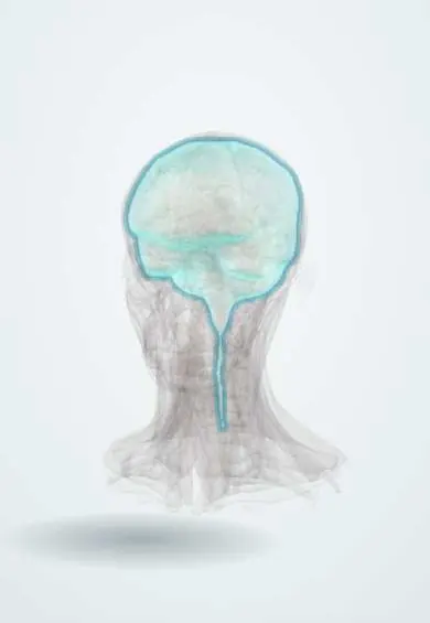
Meningioma !
A usually slow-growing, benign tumour of the brain coverings (meninges). It is the most common primary tumour inside the skull.
Updated 2026-11-01
Structures involved
Tap one to see what it is and open it in 3D.
Why it happens
- Tumour suppressor genes: RB, TP53, APC, BRCA and the two-hit hypothesis
- Benign vs malignant tumours: the comparison table
- Pathologic calcification: dystrophic vs metastatic
- Gray and white matter, cerebellar cortex, meninges
What it is
A tumour arising from arachnoid cap cells of the meninges, attached to the dura mater. Most are benign (WHO grade 1) and grow slowly, compressing rather than invading the brain.
Who gets it
The most common primary intracranial tumour in adults, peaking in middle and older age. Grade 1 tumours are about twice as common in women; atypical and anaplastic forms are more common in men. Many small meningiomas are found by chance on imaging done for other reasons.
Causes
- Arise from arachnoid cap cells of the meninges
- Loss of the NF2 gene on chromosome 22 in many tumours
- Most cases are sporadic
Risk factors
- Ionising radiation to the head, especially in childhood
- Neurofibromatosis type 2 (often multiple meningiomas)
- Female sex and hormonal factors (many express progesterone receptors)
- Older age
Pathophysiology
The tumour grows from the dura as a well-defined, extra-axial mass that pushes on the brain, cranial nerves or venous sinuses. Symptoms come from local compression, irritation of the cortex (seizures) and raised intracranial pressure. It may thicken the overlying bone (hyperostosis), and microscopy shows whorls and psammoma bodies.
Classification
- WHO grade 1: benign, slow-growing (most cases)
- WHO grade 2: atypical, higher chance of recurrence
- WHO grade 3: anaplastic (malignant), fast-growing
- By location: convexity, parasagittal and falx, sphenoid wing, olfactory groove, tuberculum sellae, posterior fossa and spinal
Symptoms and signs
- Often none; found incidentally
- Headache, sometimes worse in the morning
- Focal or generalised seizures
- Focal deficits by location: weakness of the leg (parasagittal or falx), visual loss (sphenoid wing, tuberculum sellae), loss of smell (olfactory groove)
- Personality change or apathy with frontal tumours
- Hearing loss or imbalance with posterior fossa tumours
Diagnosis
- Clinical
- Full neurological examination, including visual fields, fundoscopy and smell. Many tumours are found incidentally.
- Tests
- Histopathology after resection confirms the type and WHO grade; ophthalmological visual field testing when the optic pathways are involved.
- Imaging
- Contrast MRI is the study of choice: an extra-axial, well-defined mass with uniform enhancement and a dural tail. CT shows calcification and hyperostosis of adjacent bone.
Differential diagnosis
- Brain metastases or dural metastases
- Glioma
- Vestibular schwannoma (cerebellopontine angle)
- Pituitary adenoma (sellar region)
- Hemangiopericytoma (solitary fibrous tumour) and dural lymphoma
Goal of treatment
Relieve or prevent compression of the brain and nerves, with complete removal when safe, and long-term surveillance for regrowth.
Treatment
- Follow-up
- Small, asymptomatic tumours, especially in older adults, can be followed with serial MRI.
- Surgical
- Surgical resection, including the attached dura and any invaded bone when possible, is the main treatment for symptomatic or growing tumours.
- Procedure
- Stereotactic radiosurgery or fractionated radiotherapy for small or deep tumours, residual tumour, or grade 2–3 meningiomas.
- Medical
- Corticosteroids for symptomatic brain oedema and antiseizure drugs for patients with seizures.
Complications
- Seizures and permanent neurological deficits
- Visual loss
- Recurrence after incomplete resection or with higher grades
- Surgical complications: bleeding, infection, cerebrospinal fluid leak, venous sinus injury
Prognosis
Usually good for grade 1 tumours after complete resection. Recurrence depends on grade and extent of removal; grades 2 and 3 recur more often and have a worse outlook.
Test yourself
Think of the answer, then tap the question to see it.
What is the most common primary intracranial tumour in adults?
Meningioma.
Which MRI sign is classic for meningioma?
An extra-axial, homogeneously enhancing mass with a dural tail.
Which histological finding is typical of meningioma?
Whorled cell pattern with psammoma bodies.
Which genetic syndrome predisposes to multiple meningiomas?
Neurofibromatosis type 2 (NF2 gene, chromosome 22).
What is the main treatment for a symptomatic meningioma?
Surgical resection, with radiotherapy or radiosurgery for residual, recurrent or higher-grade tumours.
Related conditions
- Glioblastoma
- Brain metastases
- Neurofibromatosis (types 1 and 2)
- Vestibular schwannoma (acoustic neuroma)
- Pituitary adenoma
- Epilepsy and seizures
Educational information, not medical advice. For any symptom, see a health professional.
Sources: Meningioma: Diagnosis and Treatment (National Cancer Institute (NCI), 2026).
Spotted a mistake?We are always improving for the community. Tell us what is wrong and our team will review it.Report a mistake
Representative model, for educational purposes only.