Cardiovascular system · Head and neck
Anterior cerebral artery !
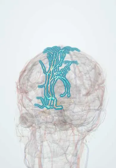
The anterior cerebral artery feeds the inner surface of the front of the brain, including the leg area; a stroke here weakens the opposite leg.
Opens in the Durgical app, no sign-up needed.
What it is
Runs forward and over the corpus callosum on the inner surface of each hemisphere.
What it does
Feeds the inner frontal and parietal lobes (leg area); a stroke here weakens the opposite leg.
Key facts
- Origin
- Internal carotid
- Branches
- Anterior communicating, pericallosal, callosomarginal, orbitofrontal
- Supplies
- Medial frontal and parietal lobes back to the parieto-occipital sulcus (leg and foot area); medial orbital surface; strip over the upper convexity; corpus callosum except the splenium; head of caudate and anterior limb of internal capsule (Heubner)
Relations
- Course
- From the end of the internal carotid it runs forward and medially above the optic nerve (A1), meets the other side at the anterior communicating artery, then climbs in the longitudinal fissure, curves round the genu of the corpus callosum and runs back over it toward the parieto-occipital sulcus
- Below
- Optic nerve and optic chiasm, below the A1 segment
- Medial
- The opposite anterior cerebral artery in the longitudinal fissure, with the falx cerebri above
How to find it
It cannot be felt. A1 runs above the optic chiasm toward the midline, and the rest of the artery curves around the genu and over the body of the corpus callosum inside the longitudinal fissure; surgeons reach it by separating the two hemispheres along the falx (interhemispheric approach).
Examination
- Grasp reflex: stroking the palm makes the fingers close and hold on involuntarily; in an adult it is a frontal release sign that fits damage to the medial frontal lobe
- Leg strength compared with arm strength on the same side (for example, the leg drifting down when held up while lying on the back): a leg clearly weaker than the arm points to this territory rather than the middle cerebral
On imaging
On a lateral angiogram it curves over the corpus callosum. On CT or MRI, an infarct in its territory is a strip along the medial frontal and parietal cortex beside the falx.
Clinical notes
- Subfalcine herniation of a swollen hemisphere can compress the anterior cerebral artery against the falx and infarct its territory
- Vasospasm after a ruptured anterior communicating aneurysm often affects both anterior cerebral arteries, giving weakness of both legs and abulia
- As in every ischaemic stroke, care is time-critical: emergency imaging and, when eligible, clot-dissolving treatment or clot retrieval
Anatomical variants
- A single (azygos) A2 feeding both hemispheres: one blockage can infarct both medial frontal lobes
- Small A1 on one side, with both distal anterior cerebral arteries filled from the other carotid
Memory aid
On the homunculus the leg hangs over the medial surface (anterior cerebral territory); the face and arm lie on the lateral surface (middle cerebral territory).
Related conditions
Test yourself
Think of the answer, then tap the question to see it.
What is the typical deficit of an anterior cerebral artery stroke?
Weakness and numbness of the opposite leg more than the arm, with the face largely spared.
Which frontal signs can follow an anterior cerebral artery infarct?
Abulia (apathy, lack of initiative), personality change, a grasp reflex and urinary incontinence.
What does the recurrent artery of Heubner supply?
The head of the caudate and the anterior limb of the internal capsule; it arises near the A1–A2 junction of the anterior cerebral artery.
Educational information, not medical advice. For any symptom, see a health professional.
Its names
- Official Latin (TA2)
- arteria anterior cerebri TA2 4502
- Official Spanish (DeCS)
- Arteria Cerebral Anterior DeCS D020771
- English (TA2)
- Anterior cerebral artery
Related
Sources: Terminologia Anatomica (TA2); DeCS; Anterior Cerebral Artery Stroke; Neuroanatomy, Circle of Willis; Tratado de anatomía humana (9.ª ed.), tomo II, Sistema nervioso central: cerebro, núcleos grises centrales, vías de conducción y circulación cerebral.
Spotted a mistake?We are always improving for the community. Tell us what is wrong and our team will review it.Report a mistake
Representative model, for educational purposes only.