Nervous system · Head and neck
Corpus callosum !
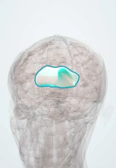
The corpus callosum is the thick arched bridge of nerve fibres joining the two halves of the brain, so they can share information.
Opens in the Durgical app, no sign-up needed.
What it is
The thick arched bridge of white matter between the two hemispheres.
What it does
Lets the two halves of the brain share information.
Key facts
- Where
- Midline floor of the longitudinal fissure
- Blood supply
- ACA (pericallosal), PCA (splenium)
- Function
- Largest commissure linking the two hemispheres
Relations
- Above
- Cingulate gyri and pericallosal arteries on each side; in the midline, the free edge of the falx cerebri and the inferior sagittal sinus
- Below
- Roof of the lateral ventricles; the septum pellucidum and the fornix hang from its underside
- In front
- Genu curving down into the rostrum, which continues to the lamina terminalis
- Behind
- Splenium over the pineal gland, the tectum and the great cerebral vein in the quadrigeminal cistern
- Lateral
- Fibres fan out into the white matter of each hemisphere; the tapetum lines the temporal and occipital horns
How to find it
Gently parting the hemispheres at the longitudinal fissure, under the falx cerebri, shows its upper surface with the pericallosal arteries running on it. On midsagittal MRI it is the thick white arch over the lateral ventricles, from the genu in front to the rounded splenium above the pineal gland and tectum.
Examination
- Tactile naming with eyes closed: after callosal section, objects placed in the left hand cannot be named while the right hand names them easily (tactile anomia)
- Gestures to spoken command with each hand: failure with the left hand only (left-sided apraxia) suggests a lesion of the front of the callosum
On imaging
Midsagittal T1 shows it as a bright arched band, thickest at the splenium. On sagittal FLAIR, multiple sclerosis plaques at the callosal–septal interface point away from the ventricles (Dawson fingers). Diffuse axonal injury often hits the splenium; a small transient splenial lesion can appear with seizures, drugs or infection.
Clinical notes
- Alexia without agraphia: a left posterior cerebral artery stroke that takes the left visual cortex and the splenium
- Marchiafava–Bignami disease: necrosis of the central callosal fibres, linked to chronic alcohol use and malnutrition
- The interhemispheric transcallosal approach opens a short slit in its body to reach the lateral and third ventricles
Development
It forms mainly between about 12 and 20 weeks of gestation, broadly from front to back with the rostrum last, so partial agenesis usually lacks the splenium and rostrum.
Related conditions
- Glycine encephalopathy (nonketotic hyperglycinaemia)
- Multiple sclerosis
- Glioblastoma
- Primary central nervous system lymphoma
- Fetal alcohol spectrum disorder
Test yourself
Think of the answer, then tap the question to see it.
What are the parts of the corpus callosum from front to back?
Rostrum, genu, body and splenium.
What happens after callosotomy when an object is felt with the left hand out of sight?
It cannot be named, because the hemispheres are disconnected.
What is a «butterfly» lesion of the corpus callosum?
A glioblastoma or lymphoma crossing the genu or splenium into both hemispheres.
What does agenesis of the corpus callosum show on imaging?
Parallel lateral ventricles, enlarged occipital horns (colpocephaly) and a high-riding third ventricle.
Educational information, not medical advice. For any symptom, see a health professional.
Its names
- Official Latin (TA2)
- corpus callosum TA2 5604
- Official Spanish (DeCS)
- Cuerpo Calloso DeCS D003337
- English (TA2)
- Corpus callosum
Related
Sources: Terminologia Anatomica (TA2); DeCS; Neuroanatomy, Corpus Callosum; 13.2 The Central Nervous System (OpenStax (Rice University), 2022) · adapted, CC BY 4.0.
Spotted a mistake?We are always improving for the community. Tell us what is wrong and our team will review it.Report a mistake
Representative model, for educational purposes only.