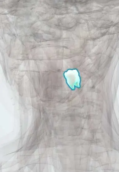
Digestive system · Head and neck
Lower left first molar !
The lower left first molar (tooth 36) is a large back tooth that comes in around age 6; it does much of the chewing.
Opens in the Durgical app, no sign-up needed.
What it is
A large back tooth with a broad chewing surface of five cusps (three on the cheek side, two on the tongue side) and two roots.
What it does
Grinds food before swallowing.
Key facts
- Joints with
- Alveolar socket of the mandible (gomphosis held by the periodontal ligament); bites against the opposing teeth
- Landmarks
- FDI 36, lower left first molar: 2 roots (mesial and distal); first permanent tooth to erupt; key to the bite (occlusion); erupts at about 6–7 years; nerve: inferior alveolar nerve (V3)
Relations
- In front
- The lower left second premolar
- Behind
- The lower left second molar
- Above
- The upper left second premolar and the upper left first molar
- Below
- Inferior alveolar canal, running below its roots inside the mandible
- Medial
- Tongue; its root tips usually lie above the mylohyoid line
How to find it
Sixth tooth from the midline on the lower left, the largest lower tooth, with five cusps; the first permanent tooth to appear, at about 6 years.
On imaging
Bitewings show decay at its contacts and in deep fissures under sealants or fillings. Periapicals show the furcation between its two roots, and a panoramic film its distance to the inferior alveolar canal.
Common problems
- Tooth decay (caries) often starts in the grooves of the chewing surface.
Clinical notes
- The area where its two roots divide (furcation) is exposed early in periodontitis and is a classic site of bone loss
- Mulberry molars, small first molars with crowded rounded cusps, are a dental sign of congenital syphilis
Anatomical variants
- A third, distolingual root (radix entomolaris) is a known variant, more frequent in people of East Asian ancestry
Development
No predecessor: it erupts behind the primary second molar. It is the first permanent tooth to begin hardening (calcifying), around birth.
Related conditions
Test yourself
Think of the answer, then tap the question to see it.
How many cusps and canals does the lower left first molar usually have?
5 cusps (3 buccal, 2 lingual) and 3 canals: 2 in the mesial root and 1, sometimes 2, in the distal root.
Why can an inferior alveolar nerve block fail to fully numb the lower left first molar?
Accessory fibres, for example from the mylohyoid nerve, may supply it; the buccal gum also needs the buccal nerve.
What is radix entomolaris?
A third, distolingual root of the lower first molar, more frequent in people of East Asian ancestry.
Why is the lower left first molar a main target for fissure sealants?
Its deep pits and fissures make it one of the teeth most prone to decay soon after it erupts.
Educational information, not medical advice. For any symptom, see a health professional.
Related
Sources: Anatomy, Head and Neck, Teeth; Anatomy, Permanent Dentition; Anatomy, Head and Neck, Tooth Eruption; Anatomy, Head and Neck: Alveolar Nerve; 23.3 The Mouth, Pharynx, and Esophagus (OpenStax (Rice University), 2022) · adapted, CC BY 4.0.
Spotted a mistake?We are always improving for the community. Tell us what is wrong and our team will review it.Report a mistake
Representative model, for educational purposes only.