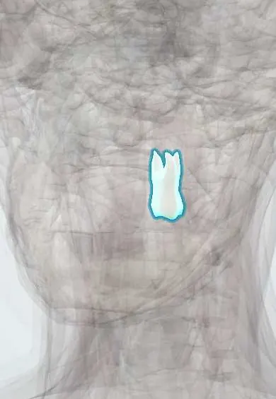
Digestive system · Head and neck
Upper left first molar !
The upper left first molar (tooth 26) is a large back tooth that comes in around age 6; it does much of the chewing.
Opens in the Durgical app, no sign-up needed.
What it is
A large back tooth with a broad chewing surface of four cusps and three roots.
What it does
Grinds food before swallowing.
Key facts
- Joints with
- Alveolar socket of the maxilla (gomphosis held by the periodontal ligament); bites against the opposing teeth
- Landmarks
- FDI 26, upper left first molar: 3 roots (2 buccal, 1 palatal); first permanent tooth to erupt; key to the bite (occlusion); erupts at about 6–7 years; nerve: posterior superior alveolar nerve (V2); roots near the floor of the maxillary sinus
Relations
- In front
- The upper left second premolar
- Behind
- The upper left second molar
- Above
- Floor of the maxillary sinus, close to all three roots
- Below
- Meets the lower left first molar and the front of the lower left second molar
How to find it
The big four-cusped tooth sixth from the midline on the upper left; it erupts at about 6 years, so in a young child it is the first tooth behind the primary molars.
On imaging
On periapical and panoramic radiographs the roots often project over the sinus floor; cone-beam CT shows whether a root actually breaks into the sinus.
Common problems
- Tooth decay (caries) often starts in the grooves of the chewing surface.
Clinical notes
- Deep decay or a failed root canal in this tooth is a common source of odontogenic sinusitis on that side; in the other direction, sinusitis can make the upper molars ache when the head is bent forward
- The area where its three roots divide (trifurcation) is a site of bone loss in periodontitis
- Its enamel can be chalky and soft from birth (molar-incisor hypomineralisation), so it breaks down and decays soon after eruption
Anatomical variants
- Cusp of Carabelli: an extra small cusp on the palatal face of the mesiopalatal cusp, seen in many populations
Development
Permanent molars grow from a backward extension of the dental lamina behind the primary teeth. This one starts to calcify around birth, so illness in early infancy can leave marks in its enamel.
Related conditions
Test yourself
Think of the answer, then tap the question to see it.
Why does the upper left first molar commonly have 4 canals?
Its mesiobuccal root often holds a second canal (MB2); missing it is a frequent cause of failed root canal treatment.
Why does decay of the upper left first molar often start early?
It replaces no tooth and erupts behind the primary second molar, so it is often mistaken for a baby tooth.
How is the upper left first molar linked to maxillary sinusitis?
Deep decay or a failed root canal in it is a common source of odontogenic sinusitis; sinusitis can also make the upper molars ache.
Why can illness in early infancy mark the enamel of the upper left first molar?
It begins to calcify around birth, so its enamel forms during the first months of life.
Educational information, not medical advice. For any symptom, see a health professional.
Related
Sources: Anatomy, Head and Neck, Teeth; Anatomy, Permanent Dentition; Anatomy, Head and Neck, Tooth Eruption; Anatomy, Head and Neck: Alveolar Nerve; 23.3 The Mouth, Pharynx, and Esophagus (OpenStax (Rice University), 2022) · adapted, CC BY 4.0.
Spotted a mistake?We are always improving for the community. Tell us what is wrong and our team will review it.Report a mistake
Representative model, for educational purposes only.