Cardiovascular system · Lower limb
Circumflex femoral veins !
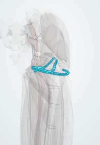
The circumflex femoral veins wrap around the top of the femur beside their arteries, draining the hip and the muscles around it.
Official name: Medial circumflex femoral veins
Opens in the Durgical app, no sign-up needed.
What it is
Veins that run with the circumflex femoral arteries around the top of the femur.
What it does
Drain the hip region into the deep femoral or femoral vein.
Key facts
- Drains
- Hip region, proximal femur and upper thigh muscles; accompany circumflex arteries
- Into
- Deep femoral vein (or femoral vein)
Relations
- Course
- The medial veins wind round the back of the femoral neck with their artery; the lateral veins come from under the sartorius and rectus femoris across the front of the hip; both end in the deep femoral vein or directly in the femoral vein
How to find it
In the direct anterior approach to the hip, the ascending lateral circumflex vessels cross the deep interval below the tensor fasciae latae and rectus femoris; the medial veins lie deeper, between pectineus and iliopsoas, then behind the femoral neck.
On imaging
On CT venography or MRI they appear as paired veins around the upper femur; when the femoral vein is blocked they may be enlarged as part of the collateral network.
Clinical notes
- In the anterior hip approach, the lateral circumflex vessels crossing the interval between the tensor fasciae latae and the sartorius are tied or sealed
- A source of bleeding in hip fractures and hip surgery
- In the anterolateral thigh flap, the paired veins of the descending branch of the lateral circumflex system are the flap's outflow and are joined to a recipient vein at the defect
Test yourself
Think of the answer, then tap the question to see it.
Where do the circumflex femoral veins drain?
Into the deep femoral vein or directly into the femoral vein, draining the hip region, upper femur and nearby muscles.
What collateral route do the circumflex femoral veins provide when the femoral vein is blocked?
They connect with the gluteal veins behind the hip, carrying thigh blood to the internal iliac vein.
Which circumflex femoral vessels are tied or sealed in the anterior hip approach?
The lateral circumflex vessels crossing the interval between the tensor fasciae latae and the sartorius.
Educational information, not medical advice. For any symptom, see a health professional.
Its names
- Official Latin (TA2)
- venae circumflexae mediales femoris; venae circumflexae laterales femoris TA2 5056
- Official Spanish (DeCS)
- Venas circunflejas femorales
- English (TA2)
- Medial circumflex femoral veins
- Also called
- venae circumflexae femoris tibiales
Related
Sources: Terminologia Anatomica (TA2); Anatomy, Abdomen and Pelvis: Femoral Triangle; 20.5 Circulatory Pathways (OpenStax (Rice University), 2022) · adapted, CC BY 4.0; Anatomy of the Human Body (20th ed.): The Arteries of the Lower Extremity (Lea & Febiger (Gray's Anatomy, 1918), 1918) · adapted, Public domain.
Spotted a mistake?We are always improving for the community. Tell us what is wrong and our team will review it.Report a mistake
Representative model, for educational purposes only.