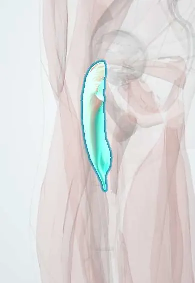
Pectineus !
The pectineus is a flat muscle at the top of the inner thigh, in the groin; it pulls the leg inward and helps lift it.
Official name: Pectineus muscle
Opens in the Durgical app, no sign-up needed.
What it is
A flat, four-sided muscle at the top of the inner thigh, from the pubic bone to just below the lesser trochanter of the femur.
What it does
Pulls the leg inward and helps bend the hip.
Key facts
- Origin
- Superior pubic ramus (pecten pubis)
- Insertion
- Pectineal line of femur, below lesser trochanter
- Action
- Adducts and flexes hip
- Nerve
- Femoral nerve (L2–L3) (varies: also a branch of obturator or accessory obturator nerve)
- Blood supply
- Medial circumflex femoral artery; obturator artery
Relations
- Superficial
- Femoral vein and femoral canal (with its lymph node), covered by the fascia lata around the saphenous opening
- Deep
- Obturator externus and adductor brevis, with the anterior branch of the obturator nerve
- Lateral
- Iliopsoas, with the medial circumflex femoral artery passing between them
- Medial
- Adductor longus
- Above
- Pecten pubis and the pectineal (Cooper) ligament
How to find it
Hard to isolate by touch: it lies in the upper femoral triangle just below the inguinal ligament, lateral to the adductor longus tendon and deep to the femoral vein. Ultrasound shows it under the femoral vessels.
On imaging
Ultrasound and MRI show it in the floor of the upper femoral triangle, deep to the femoral vein, between the iliopsoas and the adductor longus. Strains show as oedema near its origin on the pectineal line, part of the workup of athletic groin pain. A femoral hernia passes in front of it, medial to the vein, on ultrasound with Valsalva. Radiographs do not show it.
Common problems
- Groin strain: a pulled inner thigh muscle from kicking, skating, sprinting or quick changes of direction.
- Groin pain can also come from the hip joint, a hernia or the pubic bone area (athletic pubalgia), which need to be told apart.
How it is diagnosed
Physical exam: tenderness and pain when squeezing the legs together against resistance. Ultrasound or MRI for significant injuries or long-lasting pain.
Treatment
- Rest, ice, then gradual stretching and strengthening with a physical therapist.
- A planned return to sport, with hip and core strengthening to prevent repeat strains.
- Rarely, surgery for a complete tendon pull-off or long-standing groin pain that does not improve.
Clinical notes
- A femoral hernia appears below and lateral to the pubic tubercle; the repair often anchors to the pectineal (Cooper) ligament on the pecten pubis
- An obturator hernia lies hidden deep to it, which is why it is rarely felt on examination
- Femoral vein access: below the inguinal ligament the vein lies on the pectineus just medial to the femoral artery pulse
Anatomical variants
- Usually supplied by the femoral nerve, it often also gets a branch from the obturator nerve, or from an accessory obturator nerve when one is present
Development
Sits on the border between the dorsal and ventral muscle masses of the limb bud; its femoral supply with a frequent obturator contribution reflects this mixed origin, and some texts call it a composite muscle.
Test yourself
Think of the answer, then tap the question to see it.
Which triangle does the pectineus floor, and between which muscles?
The femoral triangle, between the iliopsoas laterally and the adductor longus medially.
What lies directly on the pectineus in the groin?
The femoral vein and the femoral canal; a femoral hernia comes out in front of it, below the inguinal ligament.
What is the usual nerve supply of the pectineus?
The femoral nerve (L2–L3); it may also get a branch from the obturator or accessory obturator nerve.
Educational information, not medical advice. For any symptom, see a health professional.
Its names
- Official Latin (TA2)
- musculus pectineus TA2 2627
- Official Spanish (DeCS)
- Músculo pectíneo
- English (TA2)
- Pectineus muscle
Related
- Psoas major
- Adductor brevis
- Adductor longus
- Femoral nerve
- Circumflex femoral arteries
- Deep femoral artery
- Obturator artery
- Femur
Sources: Terminologia Anatomica (TA2); Anatomy, Bony Pelvis and Lower Limb: Medial Thigh Muscles.
Spotted a mistake?We are always improving for the community. Tell us what is wrong and our team will review it.Report a mistake
Representative model, for educational purposes only.