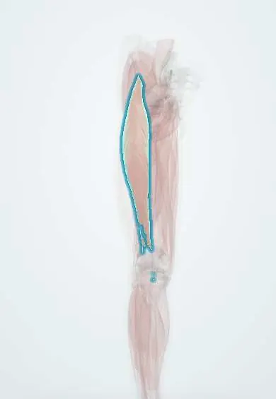
Rectus femoris !
The rectus femoris is the middle muscle of the quadriceps at the front of the thigh; it straightens the knee and lifts the thigh.
Official name: Rectus femoris muscle
Opens in the Durgical app, no sign-up needed.
What it is
The middle of the four quadriceps muscles at the front of the thigh. It is the only one that starts on the pelvis, so it crosses both the hip and the knee.
What it does
Straightens the knee and helps lift the thigh (bend the hip), as in kicking.
Key facts
- Origin
- Straight head: anterior inferior iliac spine; reflected head: ilium above acetabulum
- Insertion
- Base of patella, then tibial tuberosity via patellar ligament
- Action
- Extends knee; flexes hip
- Nerve
- Femoral nerve (L2–L4)
- Blood supply
- Lateral circumflex femoral artery (descending branch)
Relations
- Superficial
- At the top, the sartorius and the tensor fasciae latae, with the muscle appearing in the V between them; lower down only fascia lata and skin
- Deep
- Vastus intermedius over most of its length; at its origin, the front of the hip joint capsule; the descending branch of the lateral circumflex femoral artery runs under its lateral edge
- Medial
- Vastus medialis
- Lateral
- Vastus lateralis
- Below
- Joins the vasti in the quadriceps tendon above the patella, where it forms the most superficial layer
How to find it
Lying supine, the person lifts the straightened leg a little off the couch: the spindle-shaped belly stands out down the middle of the front of the thigh, from the V between the sartorius and the tensor fasciae latae to the upper border of the patella.
Examination
- Ely test: prone, the examiner bends the knee passively; if the hip flexes and the buttock rises from the couch, the rectus femoris is tight
- Modified Thomas test: with the opposite knee held to the chest and the tested thigh resting on the couch, the knee swings into extension instead of staying bent near a right angle when the rectus femoris is short
On imaging
Ultrasound or MRI show the tendon of the reflected head running down inside the muscle belly; many strains centre on this intramuscular tendon, which is described when the injury is reported.
Common problems
- Strains (pulled muscle) from sprinting or kicking; bruising from a direct blow.
- Weakness and shrinking after knee injury or surgery, which rehabilitation works to rebuild.
How it is diagnosed
Physical exam; ultrasound or MRI for significant injuries.
Treatment
- Rest, ice, then progressive strengthening with a physical therapist.
Clinical notes
- Smith-Petersen (anterior) approach to the hip: superficial interval between sartorius and tensor fasciae latae, deep interval between rectus femoris and gluteus medius; the reflected head may be released to reach the capsule
- A pedicled rectus femoris flap, based on the descending branch of the lateral circumflex femoral artery, can cover groin and lower abdominal wall defects
- With a femoral nerve lesion, knee extension fails: the knee gives way on stairs and slopes, and the patellar reflex is reduced
Anatomical variants
- A single straight head without a reflected head, and accessory slips toward the vastus lateralis, are described
Development
Like the rest of the quadriceps it comes from the dorsal muscle mass of the limb bud, hence its femoral nerve supply; the medial rotation of the lower limb in the seventh week turns this group to the front of the thigh.
Related conditions
- Myoadenylate deaminase deficiency (AMP deaminase deficiency)
- Marasmus
- Falls in older adults
- Frailty syndrome
- Sarcopenia
- Soft tissue sarcomas (liposarcoma, leiomyosarcoma, rhabdomyosarcoma)
- Inflammatory myopathies (dermatomyositis, polymyositis)
- Patellar fracture
Test yourself
Think of the answer, then tap the question to see it.
What makes the rectus femoris different from the other three quadriceps heads?
It is the only quadriceps head that crosses the hip: its straight head arises from the anterior inferior iliac spine, so it flexes the hip as well as extending the knee.
Which nerve supplies the rectus femoris, and from which roots?
The femoral nerve (L2–L4). A femoral nerve lesion makes knee extension fail, so the knee gives way on stairs, and the patellar reflex is reduced.
What does a positive Ely test show about the rectus femoris?
Tightness of the rectus femoris: prone, the knee is bent passively and the hip flexes so the buttock rises, because the muscle crosses both hip and knee.
Which avulsion can a sudden rectus femoris pull cause in an adolescent?
Avulsion of the apophysis of the anterior inferior iliac spine, the origin of the straight head of the rectus femoris.
In the Smith-Petersen anterior approach to the hip, which interval does the rectus femoris form?
The deep interval, between the rectus femoris and the gluteus medius; the superficial interval lies between the sartorius and the tensor fasciae latae.
Educational information, not medical advice. For any symptom, see a health professional.
Its names
- Official Latin (TA2)
- musculus rectus femoris TA2 2614
- Official Spanish (DeCS)
- Músculo recto femoral DeCS D052097
- English (TA2)
- Rectus femoris muscle
Related
- Vastus intermedius
- Femoral nerve
- Femoral artery
- Circumflex femoral arteries
- Rectus femoris tendon
- Hip bone
- Patella
- Patellar tendon
Sources: Terminologia Anatomica (TA2); DeCS; Anatomy, Abdomen and Pelvis, Rectus Femoris Muscle; Anatomy, Bony Pelvis and Lower Limb: Thigh Quadriceps Muscle; 8.5 Development of the Appendicular Skeleton (OpenStax (Rice University), 2022) · adapted, CC BY 4.0.
Spotted a mistake?We are always improving for the community. Tell us what is wrong and our team will review it.Report a mistake
Representative model, for educational purposes only.