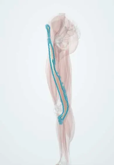
Sartorius !
The sartorius is a long strap muscle crossing the thigh from the hip to the inner knee, the longest in the body; it helps bend both joints.
Official name: Sartorius muscle
Opens in the Durgical app, no sign-up needed.
What it is
A long strap muscle crossing the front of the thigh from the hip to the inner knee.
What it does
Helps bend the hip and knee.
Key facts
- Origin
- Anterior superior iliac spine
- Insertion
- Upper medial surface of tibia (pes anserinus)
- Action
- Flexes, abducts and laterally rotates hip; flexes knee
- Nerve
- Femoral nerve (L2–L3)
- Blood supply
- Femoral artery (muscular branches)
Relations
- Course
- From the anterior superior iliac spine it spirals down and medially across the front of the thigh, passes behind the medial femoral condyle and turns forward onto the upper medial tibia
- Deep
- Upper third: iliopsoas, pectineus and the femoral nerve branches; middle third: the adductor canal with the femoral vessels, the saphenous nerve and the nerve to vastus medialis
- Medial
- Adductor longus, which meets it at the apex of the femoral triangle
- Lateral
- At its origin, the tensor fasciae latae: the two diverge from the ASIS in an inverted V, with the rectus femoris emerging between them
- Superficial
- Fascia lata and skin; at the medial knee the saphenous nerve becomes subcutaneous between its tendon and the gracilis
How to find it
Seated, with the heel of one foot slid up the opposite shin (hip flexed, abducted and laterally rotated, knee bent), the muscle stands out as a strap running from the ASIS down and inward across the thigh.
Examination
- Manual muscle test: the examiner resists the heel-up-the-opposite-shin movement; weakness points to the femoral nerve (L2–L3)
On imaging
Ultrasound and axial MRI show it as a thin strap crossing the front of the thigh from lateral to medial; in the mid-thigh it lies over the femoral vessels and saphenous nerve, the window for an adductor canal block. In adolescent athletes, an AP pelvic radiograph may show an avulsed fragment of the ASIS after a sprint or kick. MRI shows strains and pes anserinus bursitis.
Clinical notes
- Avulsion of the ASIS apophysis in adolescent sprinters and footballers, from a sudden sartorius pull
- The lateral femoral cutaneous nerve passes close to its origin, medial to the ASIS; compression there causes meralgia paresthetica
- Key landmark for exposing the femoral vessels and for the adductor canal (saphenous) nerve block used after knee surgery
- Can be turned medially as a muscle flap to cover exposed femoral vessels or a groin graft
Development
Comes from the dorsal muscle mass of the limb bud, which explains why a muscle on the inner thigh is supplied by the femoral nerve and not by the obturator nerve.
Memory aid
Pes anserinus from front to back: "Say Grace before Tea" (Sartorius, Gracilis, semiTendinosus).
Related conditions
Test yourself
Think of the answer, then tap the question to see it.
What are the attachments of the sartorius?
From the anterior superior iliac spine to the upper medial surface of the tibia (pes anserinus); it is the longest muscle in the body and crosses hip and knee.
Which borders and roofs does the sartorius form in the thigh?
The lateral border of the femoral triangle above, and the roof of the adductor canal in the middle third of the thigh.
What is the order of the pes anserinus tendons from front to back?
Sartorius, gracilis, semitendinosus («Say Grace before Tea»); the sartorius is the most anterior.
Which nerve supplies the sartorius, and how is the muscle tested?
The femoral nerve (L2–L3); the examiner resists the heel-up-the-opposite-shin movement (hip flexion, abduction and lateral rotation with the knee bent).
Educational information, not medical advice. For any symptom, see a health professional.
Its names
- Official Latin (TA2)
- musculus sartorius TA2 2610
- Official Spanish (DeCS)
- Músculo sartorio
- English (TA2)
- Sartorius muscle
Related
Sources: Terminologia Anatomica (TA2); Anatomy, Bony Pelvis and Lower Limb: Thigh Sartorius Muscle.
Spotted a mistake?We are always improving for the community. Tell us what is wrong and our team will review it.Report a mistake
Representative model, for educational purposes only.