Cardiovascular system · Lower limb
Circumflex femoral arteries !
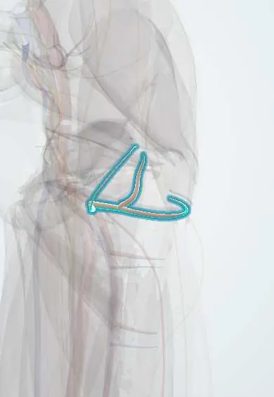
The circumflex femoral arteries are two arteries that wrap around the top of the femur; they are the main blood supply of the femoral head.
Official name: Medial circumflex femoral artery
Opens in the Durgical app, no sign-up needed.
What it is
The medial and lateral circumflex femoral arteries, usually branches of the deep femoral artery, which wrap around the top of the femur.
What it does
Supply the hip joint, the neck and head of the femur, and nearby muscles. The medial one is the main blood supply of the femoral head.
Key facts
- Origin
- Deep femoral artery (varies: either may arise directly from femoral artery)
- Branches
- Medial: retinacular arteries up femoral neck; lateral: ascending, transverse and descending branches
- Supplies
- Femoral head and neck (mainly medial circumflex), hip, adductors, quadriceps; trochanteric and cruciate anastomoses
Relations
- Course
- Medial: passes back between the pectineus and iliopsoas, then between the obturator externus and adductor brevis, and winds behind the femoral neck; lateral: runs laterally between the branches of the femoral nerve, behind the sartorius and rectus femoris, and splits into ascending, transverse and descending branches
- Behind
- The medial artery's deep branch passes behind the obturator externus tendon, then in front of the gemelli and obturator internus, close to the back of the femoral neck
- In front
- The lateral artery lies under the sartorius and rectus femoris; its ascending branch crosses the gap between the tensor fasciae latae and the sartorius
How to find it
Not palpable. In dissection of the femoral triangle the lateral circumflex runs outward among the branches of the femoral nerve, under the sartorius and rectus femoris. The medial circumflex turns backward in the gap between the pectineus and iliopsoas, then between the obturator externus and adductor brevis, heading for the back of the femoral neck.
On imaging
CT or catheter angiography shows them arising from the deep femoral artery (sometimes the femoral) and winding round the femoral neck. They are rarely imaged directly; their main role is the femoral head blood supply. MRI is the test for avascular necrosis after a neck fracture or dislocation, showing a dark line or marrow changes in the head before radiographs show collapse.
Common problems
- A femoral neck fracture or hip dislocation can damage them and cut off blood to the femoral head (avascular necrosis).
Clinical notes
- In posterior hip approaches, cutting the short external rotators too close to bone, or the obturator externus tendon, can damage the medial circumflex deep branch; techniques that keep the obturator externus intact protect the head's supply
- In the anterior hip approach, the ascending branch of the lateral circumflex vessels crosses the interval and is tied or sealed
- In children the growth plate blocks flow from the metaphysis, so the head depends on the retinacular vessels; raised joint pressure (septic arthritis, bleeding) or a slipped epiphysis can cut them off
- Hip dislocation stretches or tears the medial circumflex branches; the longer the hip stays out, the higher the risk of necrosis
Anatomical variants
- The medial circumflex femoral artery comes from the deep femoral artery in about 57% and straight from the common femoral artery in about 39%; other origins are rare
- The lateral circumflex femoral artery may arise from the common or superficial femoral artery instead of the deep femoral artery
- The ascending and descending branches of the lateral circumflex may arise separately
Related conditions
- Intertrochanteric hip fracture
- Slipped capital femoral epiphysis
- Legg-Calvé-Perthes disease
- Hip dislocation
Test yourself
Think of the answer, then tap the question to see it.
From which artery do the circumflex femoral arteries usually arise?
The deep femoral artery; either one may instead arise directly from the femoral artery.
Which circumflex femoral artery is the main blood supply of the femoral head, and through which vessels?
The medial circumflex femoral artery: its deep branch runs behind the femoral neck and gives the retinacular arteries that enter the head.
Why do displaced intracapsular femoral neck fractures risk avascular necrosis while extracapsular fractures do not?
Intracapsular displacement tears the retinacular vessels from the medial circumflex femoral artery; extracapsular fractures spare them.
Which branch of the lateral circumflex femoral artery is the pedicle of the anterolateral thigh flap?
The descending branch, running between the rectus femoris and the vastus lateralis.
Educational information, not medical advice. For any symptom, see a health professional.
Its names
- Official Latin (TA2)
- arteria circumflexa medialis femoris; arteria circumflexa lateralis femoris TA2 4686
- Official Spanish (DeCS)
- Arterias circunflejas femorales
- English (TA2)
- Medial circumflex femoral artery
- Also called
- arteria circumflexa femoris tibialis
Related
- Hip capsule
- Deep femoral artery
- Circumflex femoral veins
- Femur
- Obturator externus
- Quadratus femoris
- Pectineus
- Adductor brevis
Sources: Terminologia Anatomica (TA2); Anatomy, Bony Pelvis and Lower Limb: Lateral Circumflex Femoral Artery; Anatomy, Abdomen and Pelvis: Hip Arteries; Anatomy of the Human Body (20th ed.): The Arteries of the Lower Extremity (Lea & Febiger (Gray's Anatomy, 1918), 1918) · adapted, Public domain.
Spotted a mistake?We are always improving for the community. Tell us what is wrong and our team will review it.Report a mistake
Representative model, for educational purposes only.