Cardiovascular system · Lower limb
Femoral vein !
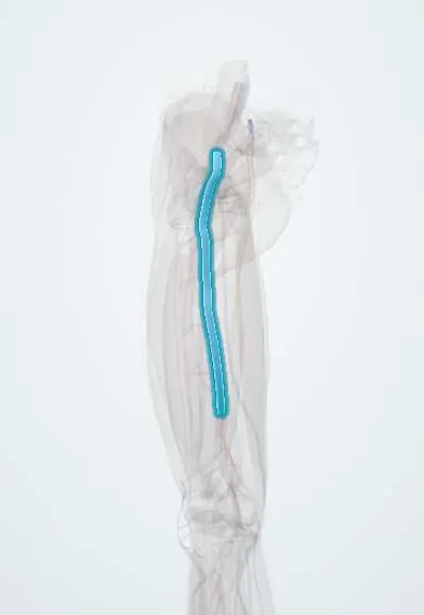
The femoral vein is the main deep vein of the thigh, beside the femoral artery; a clot in it is a deep vein thrombosis.
Opens in the Durgical app, no sign-up needed.
What it is
The main deep vein of the thigh, running beside the femoral artery from behind the knee to the groin.
What it does
Returns blood from the leg toward the heart. The pumping of the calf muscles and one-way valves help push the blood upward.
Key facts
- Drains
- Popliteal vein, deep femoral vein and great saphenous vein; medial to femoral artery at groin
- Into
- External iliac vein behind inguinal ligament
Relations
- Course
- From the adductor hiatus it rises in the adductor canal behind the artery (posterolateral to it lower down), lies behind it at the apex of the femoral triangle and medial to it at the inguinal ligament, where it becomes the external iliac vein
- Lateral
- The femoral artery; beyond it, outside the sheath, the femoral nerve
- Medial
- The femoral canal (fat, lymphatics and a lymph node), then the lacunar ligament: the femoral ring, where a femoral hernia enters; the node there is the proximal deep inguinal node (of Cloquet), which when enlarged can mimic an incarcerated femoral hernia
- In front
- Fascia lata and cribriform fascia; the great saphenous vein joins its front through the saphenous opening
- Behind
- The pectineus and, lower down, the adductor longus
How to find it
It has no pulse of its own. With the person supine and the thigh slightly turned out, the femoral pulse is found just below the inguinal ligament; the vein lies about a finger's breadth medial to it. On ultrasound it is the oval, compressible vessel medial to the round, pulsating artery.
On imaging
Compression ultrasound is the standard for DVT: in a transverse view the normal vein is medial to the artery in the groin and collapses fully under probe pressure. A clot shows as a vein that does not compress, often with echogenic material and no flow on Doppler. CT or MR venography is used for clot extending into the pelvis. Venography with contrast is now rarely needed.
Common problems
- Deep vein thrombosis (DVT): a blood clot, more likely after surgery (especially hip and knee replacement), injury, long travel, cancer or pregnancy.
- A clot can break off and travel to the lungs (pulmonary embolism), which is an emergency.
- Lasting vein damage after a clot can cause leg swelling and skin changes (post-thrombotic syndrome).
How it is diagnosed
Duplex ultrasound of the leg veins; a blood test (D-dimer); a chest CT scan if a lung clot is suspected.
Treatment
- Blood thinners (anticoagulants) and compression.
- After surgery: moving early, compression devices and sometimes medicine to prevent clots.
- For large or severe clots: procedures to remove or dissolve the clot; a vein filter in selected cases.
Clinical notes
- Femoral venous catheter: the needle enters medial to the pulse, below the inguinal ligament; these lines carry higher infection and clot rates than other sites
- A femoral hernia passes through the canal just medial to the vein; it is more common in women and prone to strangulation
Anatomical variants
- Duplication of the femoral vein in the thigh is common; on ultrasound a clot can hide in one of the two channels
- The deep femoral and circumflex femoral veins join it at variable levels
Related conditions
- Deep vein thrombosis
- Immobility syndrome and hospital-associated deconditioning
- Heparin-induced thrombocytopenia
- Inherited thrombophilia
- Antiphospholipid syndrome
Test yourself
Think of the answer, then tap the question to see it.
Is a clot in the vessel once called the «superficial femoral vein» a DVT?
Yes: it is a deep vein, now called simply the femoral vein.
Where is the femoral vein found in the groin?
Just medial to the femoral pulse, in the middle compartment of the femoral sheath, with the femoral canal medial to it.
Which superficial vein joins the femoral vein through the saphenous opening?
The great saphenous vein.
Why can a clot be missed on ultrasound of the femoral vein?
Duplication of the femoral vein in the thigh is common, and a clot can hide in one of the two channels.
Educational information, not medical advice. For any symptom, see a health professional.
Its names
- Official Latin (TA2)
- vena femoralis TA2 5055
- Official Spanish (DeCS)
- Vena femoral DeCS D005268
- English (TA2)
- Femoral vein
Related
Sources: Terminologia Anatomica (TA2); DeCS; Anatomy, Abdomen and Pelvis: Femoral Triangle; Anatomy, Abdomen and Pelvis: Femoral Sheath; Anatomía humana (4.ª ed.), tomo 1, cap. 69: Sistema linfático.
Spotted a mistake?We are always improving for the community. Tell us what is wrong and our team will review it.Report a mistake
Representative model, for educational purposes only.