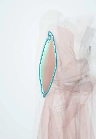
Tensor fasciae latae !
The tensor fasciae latae is a small muscle at the front-outer hip; it tightens the iliotibial band and helps steady hip and knee.
Opens in the Durgical app, no sign-up needed.
What it is
The tensor fasciae latae: a small muscle on the front-outer hip, running from the front of the pelvis into the IT band.
What it does
Helps bend the hip, move the leg out to the side and turn it inward. It keeps the IT band tight to steady the hip and knee.
Key facts
- Origin
- Anterior superior iliac spine; anterior iliac crest
- Insertion
- Iliotibial tract, which ends on lateral tibial condyle (Gerdy tubercle)
- Action
- Abducts, medially rotates and flexes hip; tenses fascia lata; steadies extended knee
- Nerve
- Superior gluteal nerve (L4–S1)
- Blood supply
- Lateral circumflex femoral artery (ascending branch); superior gluteal artery
Relations
- Medial
- Sartorius, diverging from it below the anterior superior iliac spine, with the rectus femoris appearing between them
- Behind
- Front fibres of the gluteus medius and, deeper, the gluteus minimus
- Deep
- Origin of the rectus femoris and the ascending branch of the lateral circumflex femoral artery, which enters its deep surface
- Superficial
- Fascia lata, which splits to enclose it; branches of the lateral femoral cutaneous nerve pass nearby
- Below
- Continues as the iliotibial tract down the outer thigh
How to find it
Lying supine, flexing the hip slightly and turning it inward against resistance makes a small firm belly stand out just below and behind the anterior superior iliac spine, in front of the greater trochanter.
Examination
- Ober test: lying on the unaffected side with the lower hip and knee bent, the upper leg is abducted and extended and then released; if it stays up instead of dropping toward the couch, the TFL and iliotibial tract are tight
- Thomas test: if the thigh drifts outward (abducts) as it lowers, the TFL or the iliotibial tract is short
On imaging
MRI or ultrasound shows it as a small muscle below and behind the ASIS, lateral to the sartorius, with its fibres continuing into the iliotibial tract. Look for strains or, as a late sign of superior gluteal nerve injury or gluteal tendon tears, enlargement or fatty change. The fat plane between it and the sartorius on MRI marks the direct anterior approach. Radiographs do not show it.
Common problems
- Overuse pain at the front-outer hip, often when it works too hard to make up for a weak gluteus medius.
Clinical notes
- Direct anterior hip approach: working inside the TFL sheath helps protect the lateral femoral cutaneous nerve, which lies more medially; the superior gluteal nerve branch to the muscle can be stretched
- TFL flap, based on the ascending branch of the lateral circumflex femoral artery, covers trochanteric pressure ulcers and lower abdominal wall defects
Anatomical variants
- The belly reaches below the level of the greater trochanter in about a third of people, so it may need to be split when the iliotibial tract is opened in a lateral approach
Development
Derived from the dorsal muscle mass with the gluteus medius and minimus, which explains its superior gluteal nerve even though it sits at the front of the hip, a position reached through the medial rotation of the lower limb in the embryo.
Related conditions
Test yourself
Think of the answer, then tap the question to see it.
What is the nerve supply of the tensor fasciae latae?
The superior gluteal nerve (L4–S1), so its interval with the sartorius (femoral nerve) separates two nerve territories.
Where does the tensor fasciae latae insert?
Into the iliotibial tract, which ends on the lateral tibial condyle (Gerdy tubercle).
Which test detects tightness of the tensor fasciae latae and iliotibial tract?
The Ober test: the abducted, extended upper leg stays up instead of dropping toward the couch when released.
Which artery supplies the tensor fasciae latae flap?
The ascending branch of the lateral circumflex femoral artery, which enters its deep surface.
Educational information, not medical advice. For any symptom, see a health professional.
Its names
- Official Latin (TA2)
- tensor fasciae latae TA2 2602
- Official Spanish (DeCS)
- Músculo tensor de la fascia lata
- English (TA2)
- Tensor fasciae latae
- Also called
- tensor fasciae latae muscle · tensor of fascia lata
Related
- Gluteus medius
- IT band
- Superior gluteal nerve
- Gluteal arteries
- Circumflex femoral arteries
- TFL tendon
- Hip bone
Sources: Terminologia Anatomica (TA2); Anatomy, Bony Pelvis and Lower Limb: Tensor Fasciae Latae Muscle; 8.5 Development of the Appendicular Skeleton (OpenStax (Rice University), 2022) · adapted, CC BY 4.0.
Spotted a mistake?We are always improving for the community. Tell us what is wrong and our team will review it.Report a mistake
Representative model, for educational purposes only.