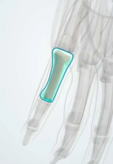
Proximal phalanx, little finger !
The proximal phalanx of the little finger is its first bone beyond the knuckle; the small muscles of the little-finger mound attach to its base.
Official name: Proximal phalanx of the little finger
Opens in the Durgical app, no sign-up needed.
What it is
The proximal bone of the little finger.
What it does
One of the finger bones; the joints between them let the finger curl.
Key facts
- Joints with
- Metacarpal head (MCP joint), next phalanx (IP joint)
- Landmarks
- Base, shaft, head (condyles)
Relations
- Above
- Head of the 5th metacarpal at the MCP joint
- In front
- FDS and FDP tendons under the A2 pulley
- Lateral
- Third palmar interosseous and the fourth lumbrical
- Medial
- Abductor digiti minimi and flexor digiti minimi
How to find it
Felt along the ulnar border of the little finger, from the fifth knuckle to the first finger joint; the hypothenar muscles reach the ulnar side of its base.
Examination
- Rotation check with the fingers flexed together: the little finger should point to the scaphoid tubercle and not cross under the ring finger
On imaging
PA and oblique views show the base and its physis in children; an extra-octave fracture appears as a Salter–Harris II line with a metaphyseal corner fragment and ulnar angulation.
Clinical notes
- Base fractures can rotate the finger: rotation is checked with the fingers flexed together
- Scarring between the shaft and the flexor tendons after a fracture can limit active bending of the little finger even when the bone heals straight
Development
Its single secondary centre at the base explains why fractures in children here cross the growth plate, as in the extra-octave fracture; the plate closes in adolescence.
Related conditions
Test yourself
Think of the answer, then tap the question to see it.
Which muscles insert on the ulnar side of the base of the proximal phalanx of the little finger?
The abductor digiti minimi and flexor digiti minimi; the third palmar interosseous inserts on the radial side.
What is an extra-octave fracture?
A Salter–Harris type II fracture of the base of the proximal phalanx of the little finger in children, with the finger deviated away from the ring finger.
How is rotation checked after a base fracture of the proximal phalanx of the little finger?
With the fingers flexed together.
Educational information, not medical advice. For any symptom, see a health professional.
Its names
- Official Latin (TA2)
- phalanx proximalis manus TA2 1277
- Official Spanish (DeCS)
- Falange proximal del meñique
- English (TA2)
- Proximal phalanx of the little finger
Related
- Digital arteries
- A1 pulleys
- Abductor digiti minimi
- Extensor tendons
- Extensor digiti minimi
- FDP tendons
- FDS tendons
- Finger joint capsules
Sources: Terminologia Anatomica (TA2); Anatomy, Shoulder and Upper Limb, Hand Bones; 8.2 Bones of the Upper Limb (OpenStax (Rice University), 2022) · adapted, CC BY 4.0; 8.5 Development of the Appendicular Skeleton (OpenStax (Rice University), 2022) · adapted, CC BY 4.0; Anatomy of the Human Body (20th ed.): Articulations of the Digits (Lea & Febiger (Gray's Anatomy, 1918), 1918) · adapted, Public domain.
Spotted a mistake?We are always improving for the community. Tell us what is wrong and our team will review it.Report a mistake
Representative model, for educational purposes only.