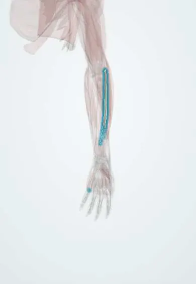
Extensor digiti minimi !
The extensor digiti minimi is a slim muscle on the back of the forearm; it lets the little finger straighten on its own.
Opens in the Durgical app, no sign-up needed.
What it is
A slim muscle that straightens the little finger.
What it does
Lets the little finger straighten on its own.
Key facts
- Origin
- Lateral epicondyle
- Insertion
- Extensor expansion of the little finger
- Action
- Extends the little finger
- Nerve
- Posterior interosseous nerve (C7–C8)
- Blood supply
- Posterior interosseous artery
Relations
- Lateral
- Extensor digitorum
- Medial
- Extensor carpi ulnaris (ECU), in the sixth compartment
- Deep
- In the forearm, extensor pollicis longus (EPL) and extensor indicis; at the wrist, the distal radioulnar joint and the ulnar head
- Course
- Through the fifth compartment, usually splitting into two slips that join the extensor mechanism of the little finger on the ulnar side of the extensor digitorum tendon
How to find it
With the other fingers curled into a fist and the little finger straightened, its tendon is felt over the back of the distal radioulnar joint, just radial to the ulnar head.
Examination
- Isolated little-finger extension with the other fingers in a fist: inability suggests a ruptured or cut tendon or posterior interosseous nerve palsy
- Wartenberg sign: the little finger rests abducted and cannot be brought to the ring finger, indicating ulnar nerve palsy
On imaging
On ultrasound its tendon, often doubled, runs alone in the fifth extensor compartment over the distal radioulnar joint. In rheumatoid arthritis an eroded ulnar head frays it, and its rupture followed by the ring and little finger extensors is the classic pattern (Vaughan-Jackson). Radiographs show the erosion; ultrasound or MRI show the tear.
Clinical notes
- Loss of little-finger extension in rheumatoid arthritis is an early warning of further tendon ruptures; treatment addresses the ulnar head as well as the tendons
- The fifth compartment is the usual window for a dorsal approach to the distal radioulnar joint
- A cut extensor tendon on the ulnar side of the back of the hand can be masked: the junctura from the ring-finger tendon still lets the little finger extend weakly, so each tendon is tested on its own
Anatomical variants
- Its tendon is commonly split in two; single, tripled or absent tendons also occur
Test yourself
Think of the answer, then tap the question to see it.
Which extensor compartment holds the extensor digiti minimi, and over what joint?
The fifth compartment, directly over the distal radioulnar joint.
Why is loss of little-finger extension important in rheumatoid arthritis?
The extensor digiti minimi is often the first tendon to rupture over a prominent ulnar head, an early warning of further ruptures (Vaughan-Jackson).
What is the Wartenberg sign, and how is the extensor digiti minimi involved?
In ulnar nerve palsy the little finger rests abducted: the extensor digiti minimi pulls unopposed by a weak third palmar interosseous.
Educational information, not medical advice. For any symptom, see a health professional.
Its names
- Official Latin (TA2)
- extensor digiti minimi TA2 2506
- Official Spanish (DeCS)
- Músculo extensor del dedo meñique
- English (TA2)
- Extensor digiti minimi
Related
Sources: Terminologia Anatomica (TA2); Anatomy, Shoulder and Upper Limb, Wrist Extensor Muscles.
Spotted a mistake?We are always improving for the community. Tell us what is wrong and our team will review it.Report a mistake
Representative model, for educational purposes only.